Institute of Neuropathology, University Hospital Erlangen, Friedrich-Alexander-University Erlangen-Nürnberg, Erlangen, Germany.
Institute of Radiology, University Hospital Erlangen, Friedrich-Alexander-University Erlangen-Nürnberg, Erlangen, Germany.
PLoS One. 2018 May 24;13(5):e0197895. doi: 10.1371/journal.pone.0197895. eCollection 2018.
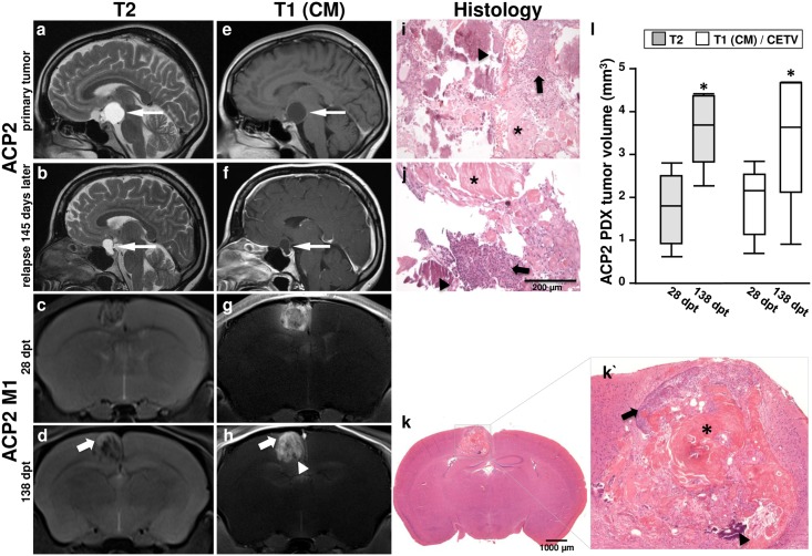
Adamantinomatous craniopharyngiomas (ACP) as benign sellar brain tumors are challenging to treat. In order to develop robust in vivo drug testing methodology, the murine orthotopic craniopharyngioma model (PDX) was characterized by magnetic resonance imaging (MRI) and histology in xenografts from three patients (ACP1-3).
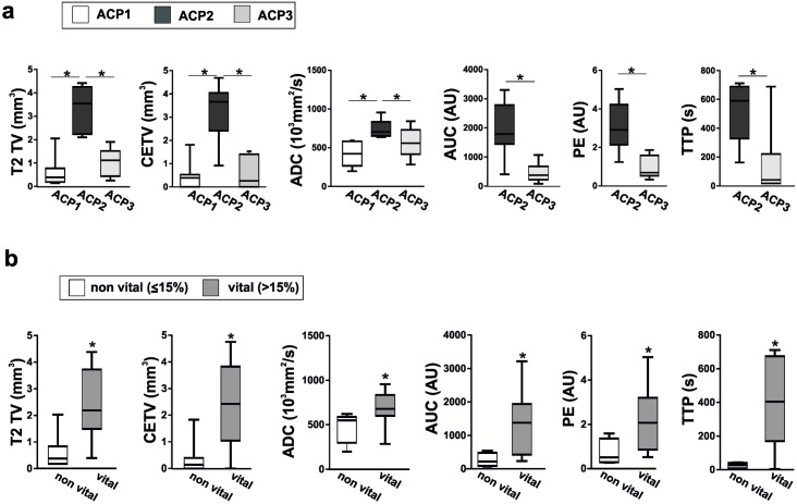
In ACP PDX, multiparametric MRI was conducted to assess morphologic characteristics such as contrast-enhancing tumor volume (CETV) as well as functional parameters from dynamic contrast-enhanced MRI (DCE-MRI) and diffusion-weighted imaging (DWI) including area-under-the-curve (AUC), peak enhancement (PE), time-to-peak (TTP) and apparent diffusion coefficient (ADC). These MRI parameters evaluated in 27 ACP PDX were correlated to histological features and percentage of vital tumor cell content.
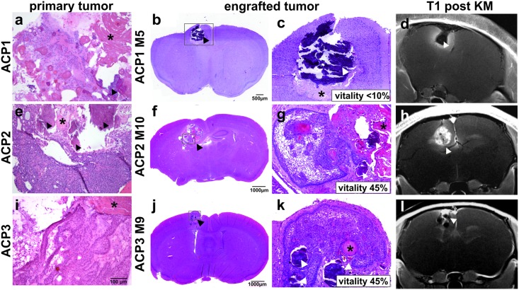
Qualitative analysis of MRI and histology from PDX revealed a similar phenotype as seen in patients, although the MRI appearance in mice resulted in a more solid tumor growth than in humans. CETV were significantly higher in ACP2 xenografts relative to ACP1 and ACP3 which correspond to respective average vitality of 41%, <10% and 26% determined histologically. Importantly, CETV prove tumor growth of ACP2 PDX as it significantly increases in longitudinal follow-up of 110 days. Furthermore, xenografts from ACP2 revealed a significantly higher AUC, PE and TTP in comparison to ACP3, and significantly increased ADC relative to ACP1 and ACP3 respectively. Overall, DCE-MRI and DWI can be used to distinguish vital from non-vital grafts, when using a cut off value of 15% for vital tumor cell content.
MRI enables the assessment of craniopharyngioma PDX vitality in vivo as validated histologically.
造釉细胞瘤型颅咽管瘤(ACP)作为良性鞍区脑肿瘤,治疗极具挑战性。为了开发稳健的体内药物测试方法,我们通过磁共振成像(MRI)和组织学对 3 名患者(ACP1-3)来源的异种移植中的鼠原位颅咽管瘤模型(PDX)进行了特征描述。
在 ACP PDX 中,进行了多参数 MRI 检查,以评估形态学特征,例如对比增强肿瘤体积(CETV),以及来自动态对比增强 MRI(DCE-MRI)和弥散加权成像(DWI)的功能参数,包括曲线下面积(AUC)、峰值增强(PE)、达峰时间(TTP)和表观弥散系数(ADC)。对 27 例 ACP PDX 进行了这些 MRI 参数评估,并与组织学特征和有活力肿瘤细胞含量的百分比相关联。
PDX 的 MRI 和组织学的定性分析显示出与患者所见相似的表型,尽管小鼠的 MRI 表现导致肿瘤生长比人类更坚实。与 ACP1 和 ACP3 相比,ACP2 异种移植中的 CETV 明显更高,这分别对应于各自组织学确定的平均活力 41%、<10%和 26%。重要的是,由于 ACP2 PDX 的 CETV 显著增加,在 110 天的纵向随访中可以证明肿瘤生长。此外,与 ACP3 相比,ACP2 的异种移植显示出明显更高的 AUC、PE 和 TTP,与 ACP1 和 ACP3 相比,ADC 分别显著增加。总体而言,当使用有活力肿瘤细胞含量 15%的截止值时,DCE-MRI 和 DWI 可用于区分有活力和无活力的移植物。
MRI 能够在体内评估颅咽管瘤 PDX 的活力,这已通过组织学验证。