文献检索文档翻译深度研究
Suppr Zotero 插件Zotero 插件
邀请有礼套餐&价格历史记录

新学期,新优惠

限时优惠:9月1日-9月22日

30天高级会员仅需29元

1天体验卡首发特惠仅需5.99元

了解详情
不再提醒
插件&应用
Suppr Zotero 插件Zotero 插件浏览器插件Mac 客户端Windows 客户端微信小程序
高级版
套餐订阅购买积分包
AI 工具
文献检索文档翻译深度研究
关于我们
关于 Suppr公司介绍联系我们用户协议隐私条款
关注我们

Suppr 超能文献

核心技术专利:CN118964589B侵权必究
粤ICP备2023148730 号-1Suppr @ 2025

半定量和定量动态对比增强(DCE)MRI 参数作为生物靶向放射治疗的前列腺癌成像生物标志物。

Semi-quantitative and quantitative dynamic contrast-enhanced (DCE) MRI parameters as prostate cancer imaging biomarkers for biologically targeted radiation therapy.

机构信息

Auckland Bioengineering Institute, The University of Auckland, Auckland, New Zealand.

School of Physics, The University of Sydney, Sydney, NSW, Australia.

出版信息

Cancer Imaging. 2022 Dec 19;22(1):71. doi: 10.1186/s40644-022-00508-9.


DOI:10.1186/s40644-022-00508-9
PMID:36536464
原文链接:https://pmc.ncbi.nlm.nih.gov/articles/PMC9762110/
Abstract

BACKGROUND: 
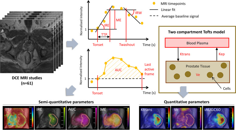
Biologically targeted radiation therapy treatment planning requires voxel-wise characterisation of tumours. Dynamic contrast enhanced (DCE) DCE MRI has shown promise in defining voxel-level biological characteristics. In this study we consider the relative value of qualitative, semi-quantitative and quantitative assessment of DCE MRI compared with diffusion weighted imaging (DWI) and T2-weighted (T2w) imaging to detect prostate cancer at the voxel level. METHODS: 
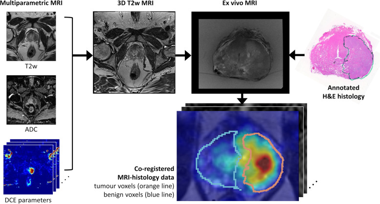
Seventy prostate cancer patients had multiparametric MRI prior to radical prostatectomy, including T2w, DWI and DCE MRI. Apparent Diffusion Coefficient (ADC) maps were computed from DWI, and semi-quantitative and quantitative parameters computed from DCE MRI. Tumour location and grade were validated with co-registered whole mount histology. Kolmogorov-Smirnov tests were applied to determine whether MRI parameters in tumour and benign voxels were significantly different. Cohen's d was computed to quantify the most promising biomarkers. The Parker and Weinmann Arterial Input Functions (AIF) were compared for their ability to best discriminate between tumour and benign tissue. Classifier models were used to determine whether DCE MRI parameters improved tumour detection versus ADC and T2w alone. RESULTS: 
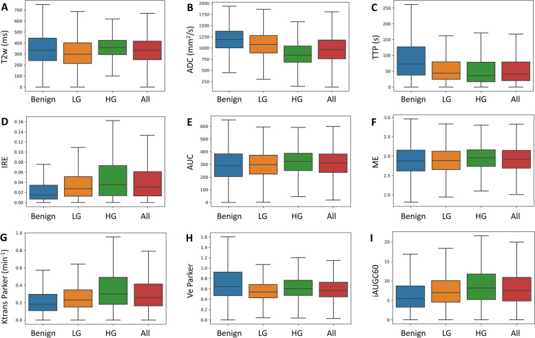
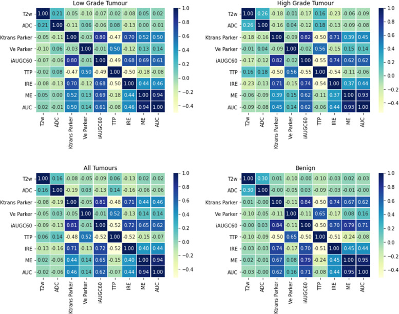
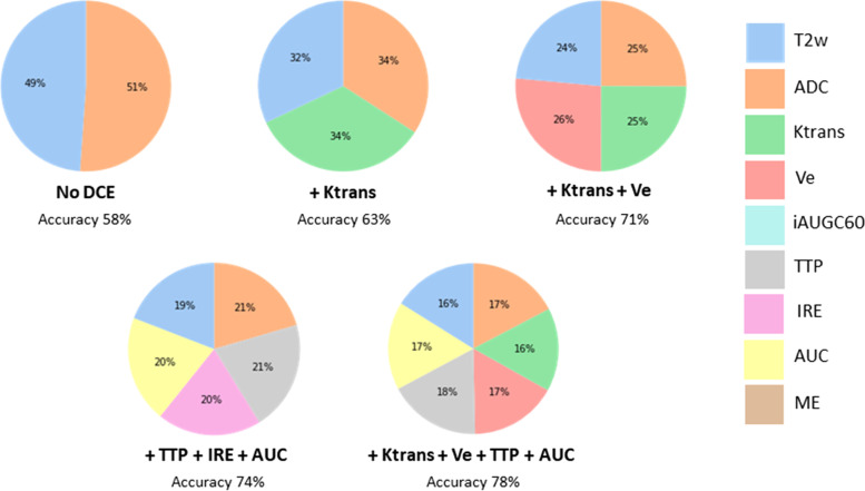
All MRI parameters had significantly different data distributions in tumour and benign voxels. For low grade tumours, semi-quantitative DCE MRI parameter time-to-peak (TTP) was the most discriminating and outperformed ADC. For high grade tumours, ADC was the most discriminating followed by DCE MRI parameters Ktrans, the initial rate of enhancement (IRE), then TTP. Quantitative parameters utilising the Parker AIF better distinguished tumour and benign voxel values than the Weinmann AIF. Classifier models including DCE parameters versus T2w and ADC alone, gave detection accuracies of 78% versus 58% for low grade tumours and 85% versus 72% for high grade tumours. CONCLUSIONS: Incorporating DCE MRI parameters with DWI and T2w gives improved accuracy for tumour detection at a voxel level. DCE MRI parameters should be used to spatially characterise tumour biology for biologically targeted radiation therapy treatment planning.

摘要

背景:生物靶向放射治疗计划需要对肿瘤进行体素级别的特征描述。动态对比增强(DCE)DCE MRI 已显示出在定义体素水平生物学特征方面的潜力。在这项研究中,我们考虑了定性、半定量和定量评估 DCE MRI 与扩散加权成像(DWI)和 T2 加权(T2w)成像相比,在体素水平检测前列腺癌的相对价值。

方法:70 例前列腺癌患者在根治性前列腺切除术前进行了多参数 MRI 检查,包括 T2w、DWI 和 DCE MRI。从 DWI 计算表观扩散系数(ADC)图,并从 DCE MRI 计算半定量和定量参数。肿瘤位置和分级通过与共配准的全切片组织学进行验证。应用 Kolmogorov-Smirnov 检验确定肿瘤和良性体素中的 MRI 参数是否存在显著差异。计算 Cohen's d 以量化最有前途的生物标志物。比较 Parker 和 Weinmann 动脉输入函数(AIF)在区分肿瘤和良性组织方面的能力。使用分类器模型确定 DCE MRI 参数是否比 ADC 和 T2w 单独提高了肿瘤检测的准确性。

结果:所有 MRI 参数在肿瘤和良性体素中的数据分布均有显著差异。对于低级别肿瘤,半定量 DCE MRI 参数达峰时间(TTP)是最具区分力的,优于 ADC。对于高级别肿瘤,ADC 是最具区分力的,其次是 DCE MRI 参数 Ktrans、初始增强率(IRE)和 TTP。利用 Parker AIF 的定量参数能更好地区分肿瘤和良性体素值,优于 Weinmann AIF。包括 DCE 参数的分类器模型与 T2w 和 ADC 单独使用相比,低级别肿瘤的检测准确率为 78%对 58%,高级别肿瘤的检测准确率为 85%对 72%。

结论:将 DCE MRI 参数与 DWI 和 T2w 结合使用,可提高体素水平肿瘤检测的准确性。DCE MRI 参数应用于空间描述肿瘤生物学,为生物靶向放射治疗计划提供依据。

https://cdn.ncbi.nlm.nih.gov/pmc/blobs/00ca/9762110/0a3858a6243e/40644_2022_508_Fig6_HTML.jpg
https://cdn.ncbi.nlm.nih.gov/pmc/blobs/00ca/9762110/713e8c80fcb8/40644_2022_508_Fig1_HTML.jpg
https://cdn.ncbi.nlm.nih.gov/pmc/blobs/00ca/9762110/742eb3ca039f/40644_2022_508_Fig2_HTML.jpg
https://cdn.ncbi.nlm.nih.gov/pmc/blobs/00ca/9762110/8d3e68dc4dbd/40644_2022_508_Fig3_HTML.jpg
https://cdn.ncbi.nlm.nih.gov/pmc/blobs/00ca/9762110/8e8f67180c9b/40644_2022_508_Fig4_HTML.jpg
https://cdn.ncbi.nlm.nih.gov/pmc/blobs/00ca/9762110/922d055bbe45/40644_2022_508_Fig5_HTML.jpg
https://cdn.ncbi.nlm.nih.gov/pmc/blobs/00ca/9762110/0a3858a6243e/40644_2022_508_Fig6_HTML.jpg

相似文献

[1]
Semi-quantitative and quantitative dynamic contrast-enhanced (DCE) MRI parameters as prostate cancer imaging biomarkers for biologically targeted radiation therapy.

Cancer Imaging. 2022-12-19

[2]
Voxel-wise correlation of positron emission tomography/computed tomography with multiparametric magnetic resonance imaging and histology of the prostate using a sophisticated registration framework.

BJU Int. 2019-1-28

[3]
Added value of diffusion-weighted images and dynamic contrast enhancement in multiparametric magnetic resonance imaging for the detection of clinically significant prostate cancer in the PICTURE trial.

BJU Int. 2019-12-11

[4]
Multiple parameters from ultrafast dynamic contrast-enhanced magnetic resonance imaging to discriminate between benign and malignant breast lesions: Comparison with apparent diffusion coefficient.

Diagn Interv Imaging. 2023-6

[5]
Prostate cancer lesion detection, volume quantification and high-grade cancer differentiation using cancer risk maps derived from multiparametric MRI with histopathology as the reference standard.

Magn Reson Imaging. 2023-6

[6]
Detecting localised prostate cancer using radiomic features in PSMA PET and multiparametric MRI for biologically targeted radiation therapy.

EJNMMI Res. 2023-4-26

[7]
Contribution of Dynamic Contrast-enhanced and Diffusion MRI to PI-RADS for Detecting Clinically Significant Prostate Cancer.

Radiology. 2023-1

[8]
A Multireader Exploratory Evaluation of Individual Pulse Sequence Cancer Detection on Prostate Multiparametric Magnetic Resonance Imaging (MRI).

Acad Radiol. 2018-4-25

[9]
Utility of Quantitative T2-Mapping Compared to Conventional and Advanced Diffusion Weighted Imaging Techniques for Multiparametric Prostate MRI in Men with Hip Prosthesis.

J Magn Reson Imaging. 2022-1

[10]
What is the most effective tool for detecting prostate cancer using a standard MR scanner?

Magn Reson Med Sci. 2013-10-29

引用本文的文献

[1]
Risk prediction models for biochemical recurrence of Chinese prostate cancer patients after radical prostatectomy based on magnetic resonance imaging examination: a systematic review.

Quant Imaging Med Surg. 2025-9-1

[2]
Analysis of dynamic contrast-enhanced T1-weighted imaging parameters in type II TIC breast lesions.

Front Oncol. 2025-8-12

[3]
The Role of Dynamic Contrast Enhanced Magnetic Resonance Imaging in Evaluating Prostate Adenocarcinoma: A Partially-Blinded Retrospective Study of a Prostatectomy Patient Cohort With Whole Gland Histopathology Correlation and Application of PI-RADS or TNM Staging.

Prostate. 2025-4

[4]
Advantage of whole-mount histopathology in prostate cancer: current applications and future prospects.

BMC Cancer. 2024-4-11

[5]
Focal Boost in Prostate Cancer Radiotherapy: A Review of Planning Studies and Clinical Trials.

Cancers (Basel). 2023-10-8

[6]
Quantitative Dynamic Contrast-Enhanced Magnetic Resonance Parameters Could Predict International Society of Urological Pathology Risk Groups of Prostate Cancers on Radical Prostatectomy.

Life (Basel). 2023-9-21

本文引用的文献

[1]
The current role of MRI for guiding active surveillance in prostate cancer.

Nat Rev Urol. 2022-6

[2]
Biologically Targeted Radiation Therapy: Incorporating Patient-Specific Hypoxia Data Derived from Quantitative Magnetic Resonance Imaging.

Cancers (Basel). 2021-9-29

[3]
Impact of qualitative, semi-quantitative, and quantitative analyses of dynamic contrast-enhanced magnet resonance imaging on prostate cancer detection.

PLoS One. 2021

[4]
Dynamic Contrast-Enhanced MRI of Prostate Lesions of Simultaneous [Ga]Ga-PSMA-11 PET/MRI: Comparison between Intraprostatic Lesions and Correlation between Perfusion Parameters.

Cancers (Basel). 2021-3-19

[5]
Biparametric versus multiparametric magnetic resonance imaging of the prostate: detection of clinically significant cancer in a perfect match group.

Prostate Int. 2020-12

[6]
Quantitative MRI: Defining repeatability, reproducibility and accuracy for prostate cancer imaging biomarker development.

Magn Reson Imaging. 2021-4

[7]
Is dynamic contrast enhancement still necessary in multiparametric magnetic resonance for diagnosis of prostate cancer: a systematic review and meta-analysis.

Transl Androl Urol. 2020-4

[8]
Can Biparametric Prostate Magnetic Resonance Imaging Fulfill its PROMIS?

Eur Urol. 2020-10

[9]
Progress towards Patient-Specific, Spatially-Continuous Radiobiological Dose Prescription and Planning in Prostate Cancer IMRT: An Overview.

Cancers (Basel). 2020-4-1

[10]
Arguments against using an abbreviated or biparametric prostate MRI protocol.

Abdom Radiol (NY). 2020-12

文献AI研究员

20分钟写一篇综述,助力文献阅读效率提升50倍

立即体验

用中文搜PubMed

大模型驱动的PubMed中文搜索引擎

马上搜索

推荐工具

医学文档翻译智能文献检索