Centre for Neuroimaging Sciences, King's College London, London, United Kingdom.
The Computational, Cognitive and Clinical Neuroimaging Laboratory, The Centre for Neuroscience, The Division of Brain Sciences, Imperial College London, Hammersmith Hospital Campus, London, United Kingdom.
PLoS One. 2018 May 24;13(5):e0197893. doi: 10.1371/journal.pone.0197893. eCollection 2018.
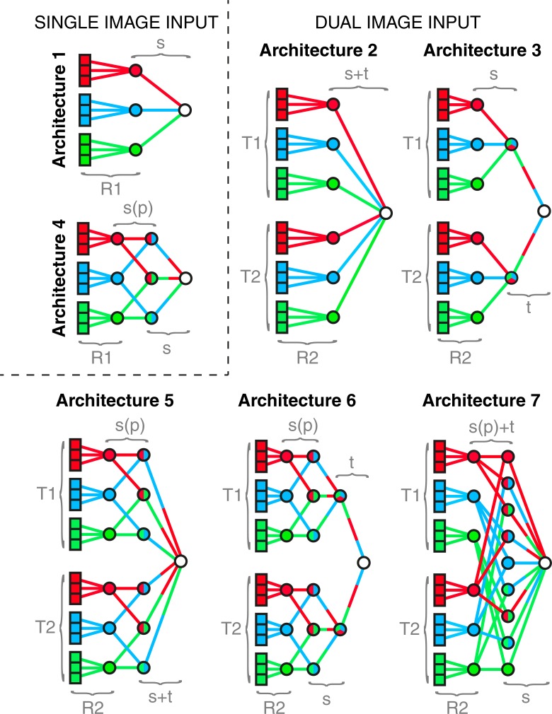
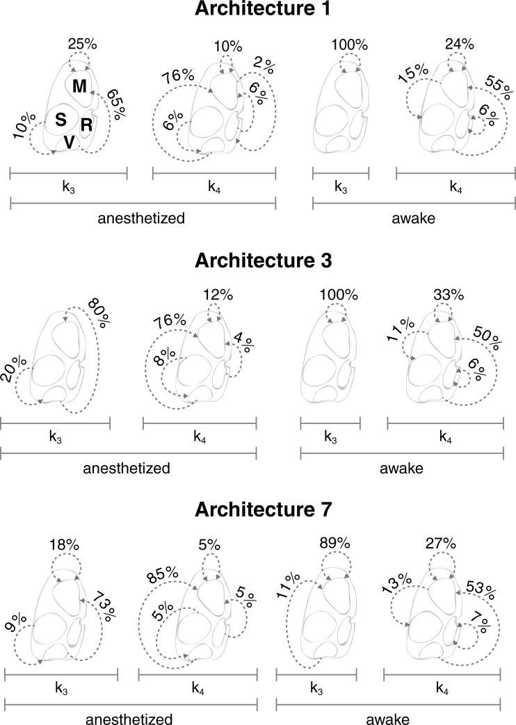
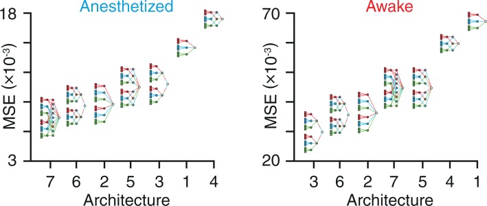
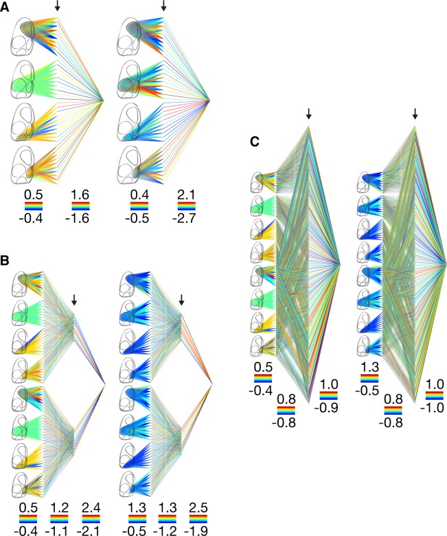
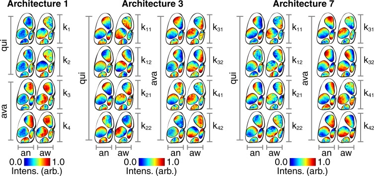
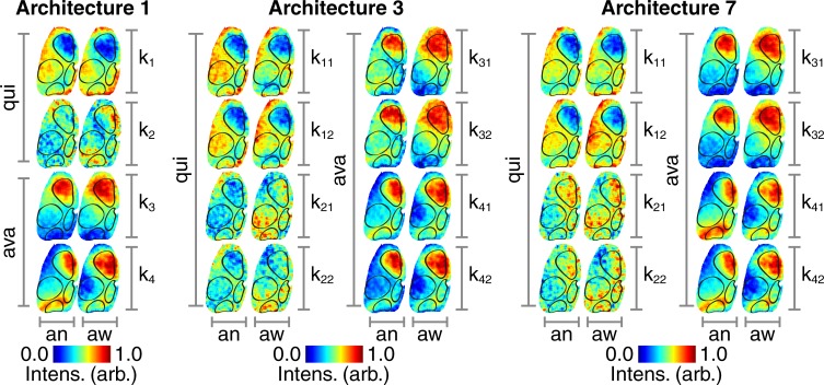
Local perturbations within complex dynamical systems can trigger cascade-like events that spread across significant portions of the system. Cascades of this type have been observed across a broad range of scales in the brain. Studies of these cascades, known as neuronal avalanches, usually report the statistics of large numbers of avalanches, without probing the characteristic patterns produced by the avalanches themselves. This is partly due to limitations in the extent or spatiotemporal resolution of commonly used neuroimaging techniques. In this study, we overcome these limitations by using optical voltage (genetically encoded voltage indicators) imaging. This allows us to record cortical activity in vivo across an entire cortical hemisphere, at both high spatial (30um) and temporal (20ms) resolution in mice that are either in an anesthetized or awake state. We then use artificial neural networks to identify the characteristic patterns created by neuronal avalanches in our data. The avalanches in the anesthetized cortex are most accurately classified by an artificial neural network architecture that simultaneously connects spatial and temporal information. This is in contrast with the awake cortex, in which avalanches are most accurately classified by an architecture that treats spatial and temporal information separately, due to the increased levels of spatiotemporal complexity. This is in keeping with reports of higher levels of spatiotemporal complexity in the awake brain coinciding with features of a dynamical system operating close to criticality.
局部扰动会在复杂动力系统中引发级联事件,这些事件会在系统的重要部分传播。这种级联事件在大脑的广泛范围内都有观察到。对这些级联事件(称为神经元瀑流)的研究通常报告大量瀑流的统计数据,而没有探测瀑流本身产生的特征模式。这部分是由于常用神经影像学技术的范围或时空分辨率的限制。在这项研究中,我们使用光学电压(遗传编码电压指示剂)成像来克服这些限制。这使我们能够在麻醉或清醒状态下的小鼠中,以高空间(约 30μm)和高时间(约 20ms)分辨率,在整个皮质半球上记录皮质活动。然后,我们使用人工神经网络来识别我们数据中神经元瀑流产生的特征模式。麻醉皮质中的瀑流可以通过同时连接空间和时间信息的人工神经网络架构进行最准确的分类。这与清醒皮质形成对比,在清醒皮质中,由于时空复杂性的增加,空间和时间信息分别处理的架构可以对瀑流进行最准确的分类。这与报告中的清醒大脑的时空复杂性水平更高一致,这与接近临界状态的动力系统的特征一致。