Setiohadji Bambang, Irfani Irawati, Rifada Maula, Virgana Rova, Kartasasmita Arief S
Ophthalmology Department, Universitas Padjadjaran, Bandung, Indonesia.
Cicendo National Eye Hospital, Bandung, Indonesia.
Ophthalmol Ther. 2018 Jun;7(1):167-172. doi: 10.1007/s40123-018-0132-z. Epub 2018 May 24.
The incidence of blindness due to methanol intoxication is higher in males of productive age. The management of methanol-induced toxic optic neuropathy is yet to produce satisfactory results. Antioxidant therapy is now used as an alternative method of preventing methanol intoxication. The aim of this study was to observe the effect of TEMPOL (4-hydroxy-2,2,6,6-tetramethylpiperidinyl-1-oxyl), a superoxide dismutase (SOD) mimetic, on retinal ganglion cells in methanol-intoxicated rats.
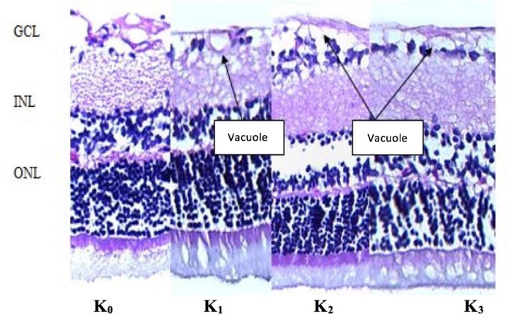
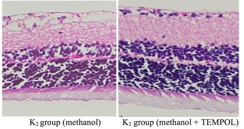
This experimental study was conducted with 20 male Wistar rats that were 10-12 weeks old and weighed 300-350 g. The rats were divided into four groups that each received a different treatment: a negative control group, a positive control group, a methanol group, and a methanol + TEMPOL group. Enucleated eyes from all groups were sliced and stained using hematoxylin-eosin (HE). Retinal layer and ganglion cells were assessed based on cellular structure, cellular swelling, and vacuole formation in the ganglion cell layer as observed at × 200 magnification. The Kruskal-Wallis test and the Mann-Whitney test were used, with significance taken to correspond to p < 0.05.
Retinal ganglion cells of the control group had fewer vacuoles and a more well-organized cellular structure compared to those of the methanol group. The histopathologic scores of the methanol-intoxicated group were lower than those of the TEMPOL therapy group; p = 0.011 (i.e., p < 0.05).
TEMPOL had a positive impact on the cellular structure of retinal ganglion cells in methanol-intoxicated rats.
在育龄男性中,甲醇中毒导致失明的发生率较高。甲醇中毒性视神经病变的治疗尚未取得令人满意的效果。抗氧化疗法现被用作预防甲醇中毒的替代方法。本研究的目的是观察超氧化物歧化酶(SOD)模拟物TEMPOL(4-羟基-2,2,6,6-四甲基哌啶-1-氧基)对甲醇中毒大鼠视网膜神经节细胞的影响。
本实验研究使用了20只10 - 12周龄、体重300 - 350克的雄性Wistar大鼠。将大鼠分为四组,每组接受不同的处理:阴性对照组、阳性对照组、甲醇组和甲醇+TEMPOL组。对所有组的摘除眼球进行切片,并用苏木精-伊红(HE)染色。在200倍放大倍数下观察,根据神经节细胞层的细胞结构、细胞肿胀和液泡形成情况评估视网膜层和神经节细胞。使用Kruskal-Wallis检验和Mann-Whitney检验,显著性水平设定为p < 0.05。
与甲醇组相比,对照组的视网膜神经节细胞液泡较少,细胞结构更有序。甲醇中毒组的组织病理学评分低于TEMPOL治疗组;p = 0.011(即p < 0.05)。
TEMPOL对甲醇中毒大鼠视网膜神经节细胞的细胞结构有积极影响。