Department of Orthopedics, The Second Affiliated Hospital of Harbin Medical University, Harbin, Heilongjiang Province 150086, China.
Departmentof Pharmacology (the State-Province Key Laboratories of Biomedicine-Pharmaceutics of China), Harbin Medical University, Harbin, eilongjiang Province 150081, China.
Int J Biol Sci. 2018 Apr 5;14(5):497-507. doi: 10.7150/ijbs.22409. eCollection 2018.
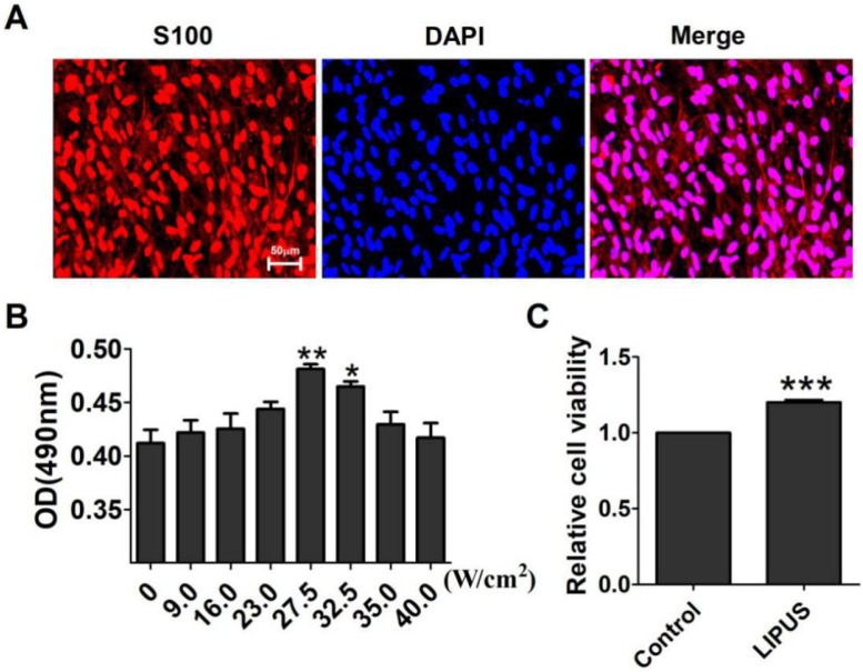
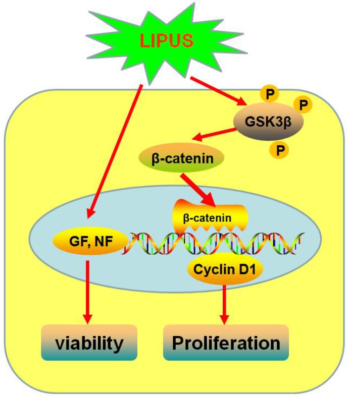
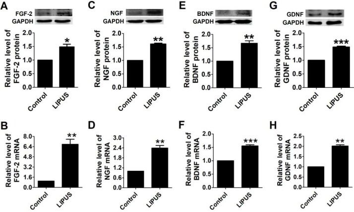
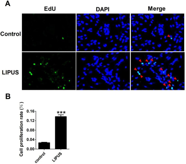
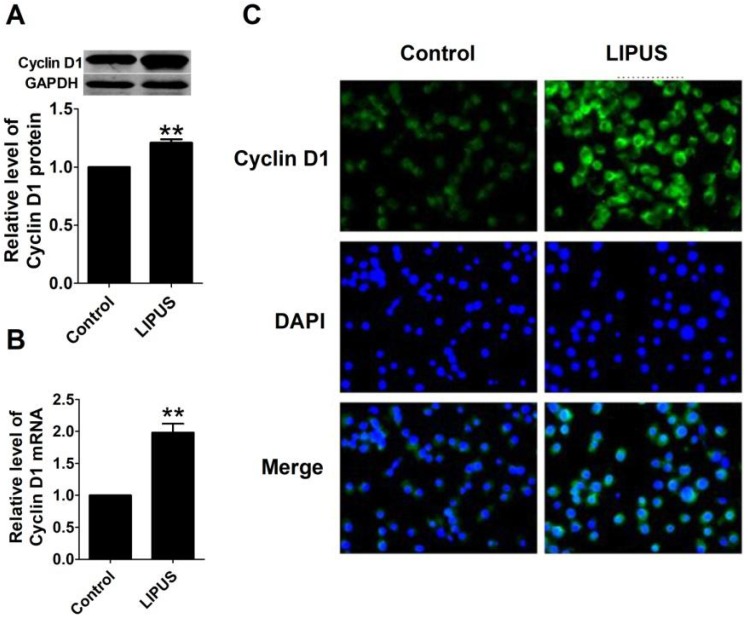
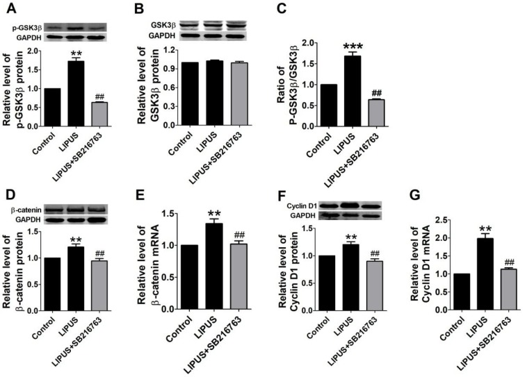
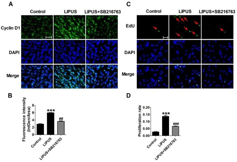
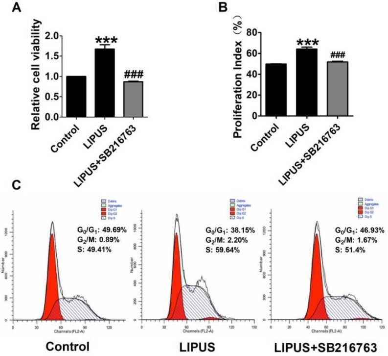
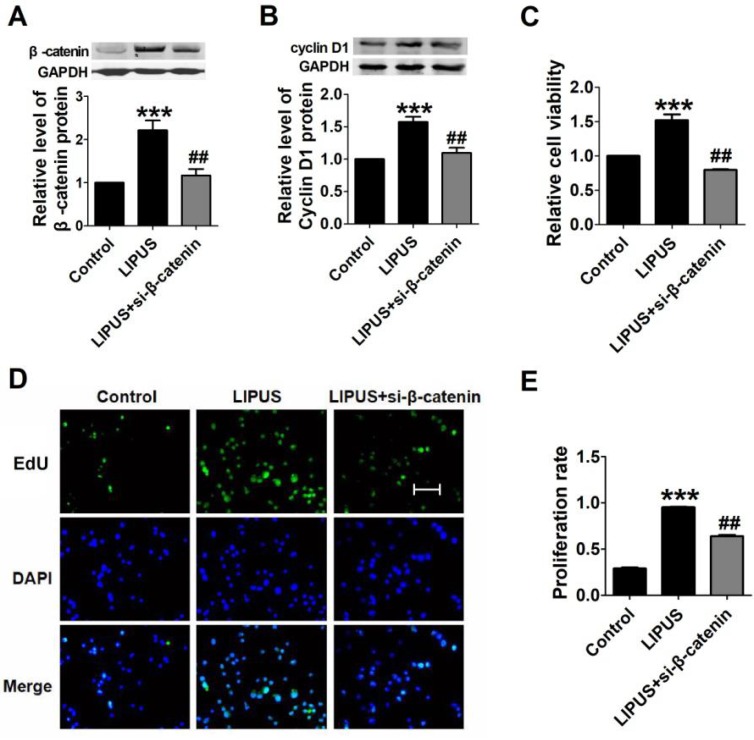
: It has been reported that ultrasound enhances peripheral nerve regeneration, but the mechanism remains elusive. Low-intensity pulsed ultrasound (LIPUS) has been reported to enhance proliferation and alter protein production in various types of cells. In this study, we detected the effects of LIPUS on Schwann cells. : Schwann cells were separated from new natal Sprague-Dawley rat sciatic nerves and were cultured and purified. The Schwann cells were treated by LIPUS for 10 minutes every day, with an intensity of 27.37 mW/cm. After treatment for 5 days, MTT, EdU staining, and flow cytometry were performed to examine cell viability and proliferation. Neurotrophic factors, including FGF, NGF, BDNF, and GDNF, were measured by western blot and real-time PCR. GSK-3β, p-GSK-3β, β-catenin and Cyclin D1 protein levels were detected using a western blot analysis. The expression of Cyclin D1 was also detected by immunofluorescence. : MTT and EdU staining showed that LIPUS increased the Schwann cells viability and proliferation. Compared to the control group, LIPUS increased the expression of growth factors and neurotrophic factors, including FGF, NGF, BDNF, GDNF, and Cyclin D1. Meanwhile, GSK-3β activity was inhibited in the LIPUS group as demonstrated by the increased level of p-GSK-3β and the ratio of the p-GSK-3β/GSK-3β level. The mRNA and protein expressions of β-catenin were increased in the LIPUS group. However, SB216763, a GSK-3β inhibitor, reversed the effects of LIPUS on Schwann cells. : LIPUS promotes Schwann cell viability and proliferation by increasing Cyclin D1 expression via enhancing the GSK-3β/β-catenin signaling pathway.
已有报道称超声可促进周围神经再生,但具体机制尚不清楚。低强度脉冲超声(LIPUS)已被报道可促进多种类型细胞的增殖和改变蛋白产物的生成。在本研究中,我们检测了 LIPUS 对许旺细胞的影响。
许旺细胞从新生 Sprague-Dawley 大鼠坐骨神经中分离出来并进行培养和纯化。LIPUS 每天处理 Schwann 细胞 10 分钟,强度为 27.37 mW/cm。处理 5 天后,通过 MTT、EdU 染色和流式细胞术检测细胞活力和增殖。通过 Western blot 和实时 PCR 测定神经营养因子,包括 FGF、NGF、BDNF 和 GDNF。通过 Western blot 分析检测 GSK-3β、p-GSK-3β、β-catenin 和 Cyclin D1 蛋白水平。通过免疫荧光检测 Cyclin D1 的表达。
MTT 和 EdU 染色表明 LIPUS 增加了 Schwann 细胞的活力和增殖。与对照组相比,LIPUS 增加了生长因子和神经营养因子的表达,包括 FGF、NGF、BDNF、GDNF 和 Cyclin D1。同时,LIPUS 组中 GSK-3β 的活性受到抑制,表现为 p-GSK-3β 水平增加和 p-GSK-3β/GSK-3β 水平的比值增加。LIPUS 组中β-catenin 的 mRNA 和蛋白表达增加。然而,GSK-3β 抑制剂 SB216763 逆转了 LIPUS 对 Schwann 细胞的作用。
LIPUS 通过增强 GSK-3β/β-catenin 信号通路增加 Cyclin D1 的表达,从而促进 Schwann 细胞的活力和增殖。