Department of Experimental Medical Science, Experimental Neuroinflammation Laboratory, Lund University, Lund, Sweden.
Department of Biochemistry and Structural Biology, Lund University, Lund, Sweden.
J Neuroinflammation. 2018 May 28;15(1):168. doi: 10.1186/s12974-018-1204-7.
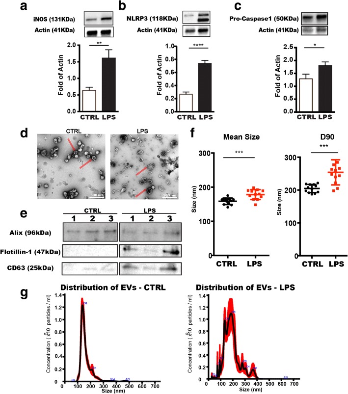
Activated microglia play an essential role in inflammatory responses elicited in the central nervous system (CNS). Microglia-derived extracellular vesicles (EVs) are suggested to be involved in propagation of inflammatory signals and in the modulation of cell-to-cell communication. However, there is a lack of knowledge on the regulation of EVs and how this in turn facilitates the communication between cells in the brain. Here, we characterized microglial EVs under inflammatory conditions and investigated the effects of inflammation on the EV size, quantity, and protein content.
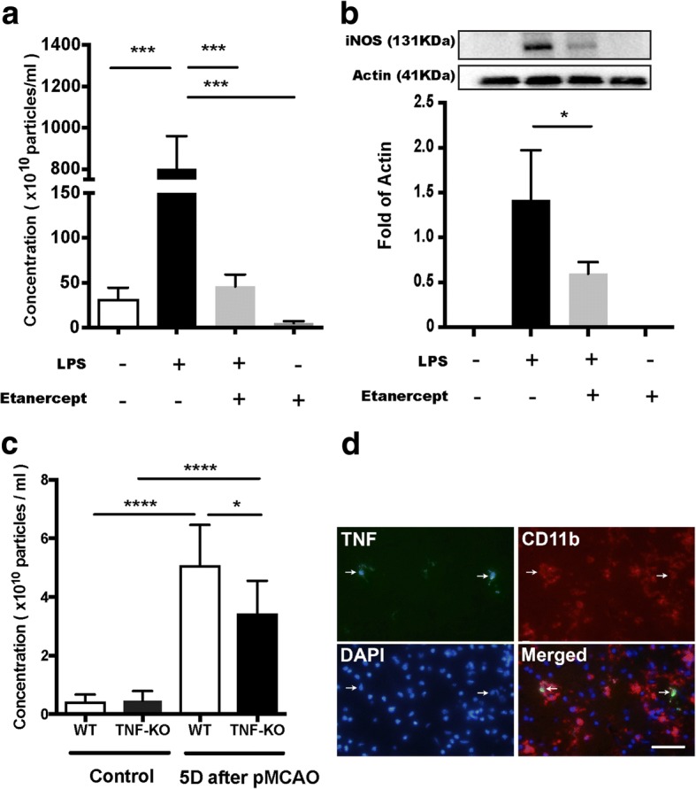
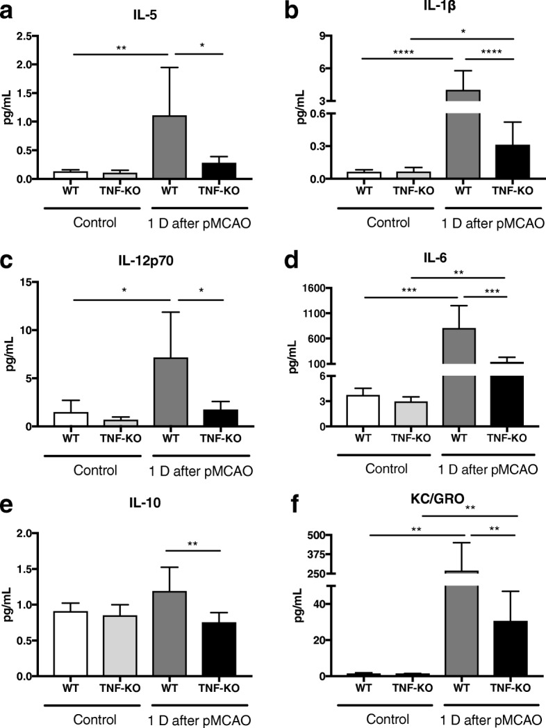
We have utilized western blot, nanoparticle tracking analysis (NTA), and mass spectrometry to characterize EVs and examine the alterations of secreted EVs from a microglial cell line (BV2) following lipopolysaccharide (LPS) and tumor necrosis factor (TNF) inhibitor (etanercept) treatments, or either alone. The inflammatory responses were measured with multiplex cytokine ELISA and western blot. We also subjected TNF knockout mice to experimental stroke (permanent middle cerebral artery occlusion) and validated the effect of TNF inhibition on EV release.
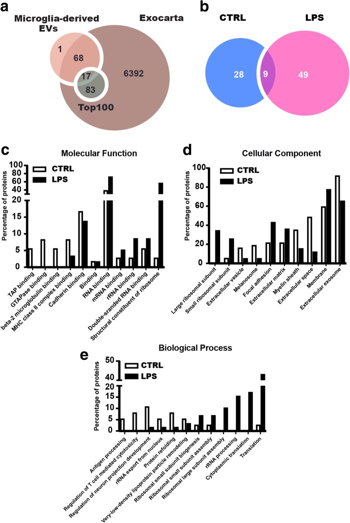
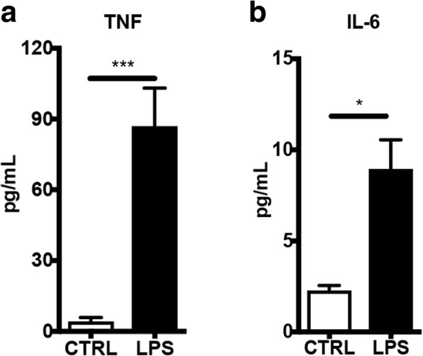
Our analysis of EVs originating from activated BV2 microglia revealed a significant increase in the intravesicular levels of TNF and interleukin (IL)-6. We also observed that the number of EVs released was reduced both in vitro and in vivo when inflammation was inhibited via the TNF pathway. Finally, via mass spectrometry, we identified 49 unique proteins in EVs released from LPS-activated microglia compared to control EVs (58 proteins in EVs released from LPS-activated microglia and 37 from control EVs). According to Gene Ontology (GO) analysis, we found a large increase of proteins related to translation and transcription in EVs from LPS. Importantly, we showed a distinct profile of proteins found in EVs released from LPS treated cells compared to control.
We demonstrate altered EV production in BV2 microglial cells and altered cytokine levels and protein composition carried by EVs in response to LPS challenge. Our findings provide new insights into the potential roles of EVs that could be related to the pathogenesis in neuroinflammatory diseases.
激活的小胶质细胞在中枢神经系统(CNS)中引发的炎症反应中发挥着重要作用。小胶质细胞衍生的细胞外囊泡(EVs)被认为参与炎症信号的传播,并调节细胞间的通讯。然而,人们对 EV 的调节以及这反过来如何促进大脑细胞间的通讯知之甚少。在这里,我们在炎症条件下对小胶质细胞 EV 进行了表征,并研究了炎症对 EV 大小、数量和蛋白含量的影响。
我们利用 Western blot、纳米颗粒跟踪分析(NTA)和质谱法来表征 EVs,并研究脂多糖(LPS)和肿瘤坏死因子(TNF)抑制剂(依那西普)处理或单独处理后,源自小胶质细胞系(BV2)的分泌 EV 的变化。通过多重细胞因子 ELISA 和 Western blot 测量炎症反应。我们还使 TNF 敲除小鼠发生实验性中风(永久性大脑中动脉闭塞),并验证了 TNF 抑制对 EV 释放的影响。
我们对源自激活的 BV2 小胶质细胞的 EV 的分析显示,TNF 和白细胞介素(IL)-6 的囊内水平显著增加。我们还观察到,当通过 TNF 途径抑制炎症时,无论是在体外还是体内,释放的 EV 数量都减少了。最后,通过质谱分析,我们发现与对照 EV 相比,LPS 激活的小胶质细胞释放的 EV 中有 49 种独特的蛋白质(LPS 激活的小胶质细胞释放的 EV 中有 58 种蛋白质,对照 EV 中有 37 种蛋白质)。根据基因本体论(GO)分析,我们发现 LPS 中与翻译和转录相关的蛋白质大量增加。重要的是,我们发现与对照相比,LPS 处理细胞释放的 EV 中存在明显不同的蛋白质谱。
我们证明了 LPS 刺激后 BV2 小胶质细胞中 EV 的产生以及 EV 中细胞因子水平和蛋白组成发生改变。我们的研究结果提供了有关 EV 可能与神经炎症性疾病发病机制相关的潜在作用的新见解。