California Institute of Technology, Pasadena, CA 91125.
Howard Hughes Medical Institute, Chevy Chase, MD 20815.
Mol Biol Cell. 2018 Jun 1;29(11):1318-1331. doi: 10.1091/mbc.E17-12-0736. Epub 2018 Apr 10.
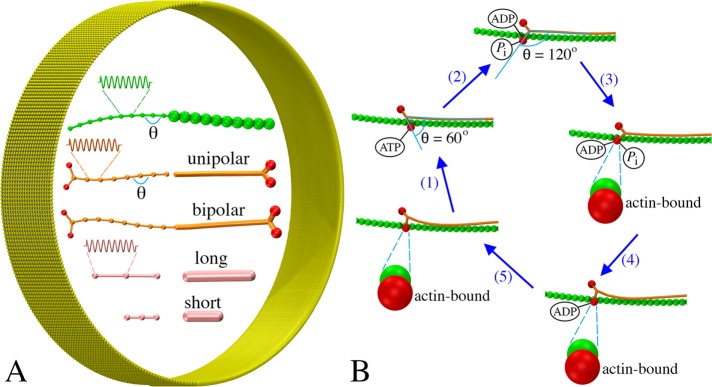
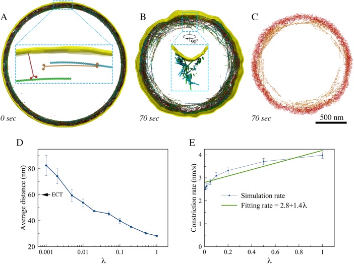
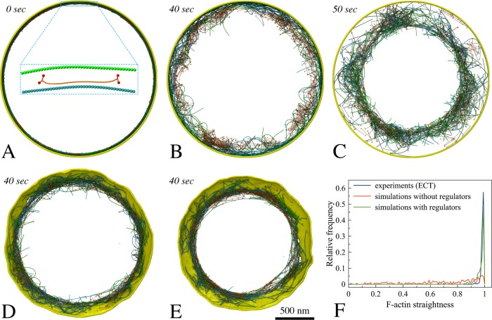
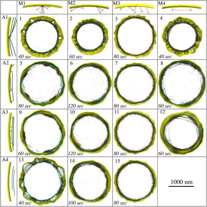
Cytokinesis in many eukaryotic cells is orchestrated by a contractile actomyosin ring. While many of the proteins involved are known, the mechanism of constriction remains unclear. Informed by the existing literature and new three-dimensional (3D) molecular details from electron cryotomography, here we develop 3D coarse-grained models of actin filaments, unipolar and bipolar myosins, actin cross-linkers, and membranes and simulate their interactions. Assuming that local force on the membrane results in inward growth of the cell wall, we explored a matrix of possible actomyosin configurations and found that node-based architectures like those presently described for ring assembly result in membrane puckers not seen in electron microscope images of real cells. Instead, the model that best matches data from fluorescence microscopy, electron cryotomography, and biochemical experiments is one in which actin filaments transmit force to the membrane through evenly distributed, membrane-attached, unipolar myosins, with bipolar myosins in the ring driving contraction. While at this point this model is only favored (not proven), the work highlights the power of coarse-grained biophysical simulations to compare complex mechanistic hypotheses.
在许多真核细胞中,胞质分裂是由收缩的肌动球蛋白环来协调的。尽管已经知道许多相关的蛋白质,但收缩的机制仍不清楚。根据现有文献和电子断层扫描的新三维(3D)分子细节,我们在这里开发了肌动蛋白丝、单极和双极肌球蛋白、肌动蛋白交联蛋白和膜的 3D 粗粒模型,并模拟了它们的相互作用。假设膜上的局部力导致细胞壁向内生长,我们探索了一组可能的肌球蛋白构型,发现像目前描述的环组装那样基于节点的结构会导致膜起皱,而不是在真实细胞的电子显微镜图像中看到的情况。相反,与荧光显微镜、电子断层扫描和生化实验数据最匹配的模型是,肌动蛋白丝通过均匀分布的、附着在膜上的单极肌球蛋白将力传递到膜上,而环中的双极肌球蛋白则驱动收缩。虽然目前这种模型只是更受青睐(而非被证明),但这项工作突出了粗粒生物物理模拟在比较复杂的机械假说方面的作用。