Erinanc Hilal, Türk Emin
Medicine Faculty, Pathology Department, Konya Uygulama ve Arastırma Hastanesi, Baskent University, Selcuklu, Konya, Turkey.
Medicine Faculty, Surgery Department, Konya Uygulama ve Arastırma Hastanesi, Baskent University, Selcuklu, Konya, Turkey.
Case Rep Pathol. 2018 Apr 30;2018:1612587. doi: 10.1155/2018/1612587. eCollection 2018.
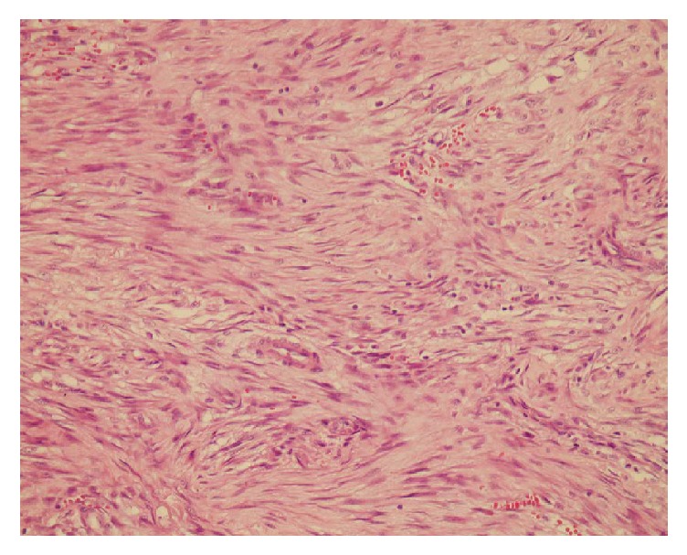
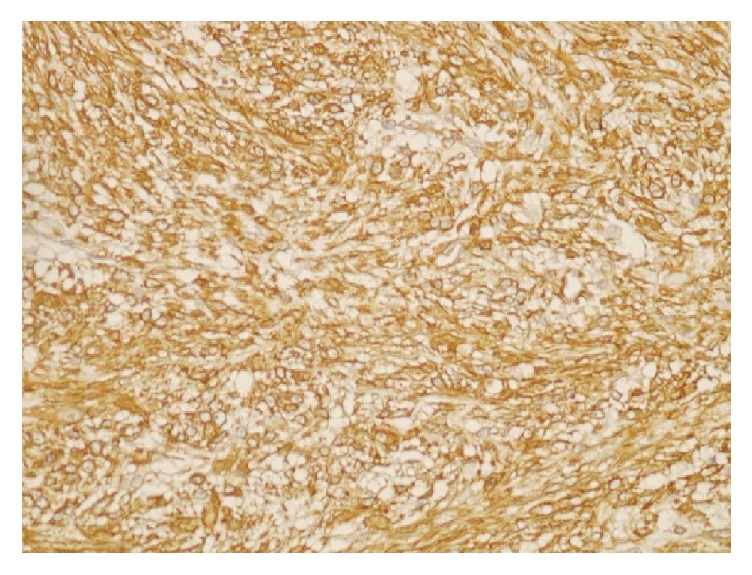
We herein report the clinical and pathological findings of a rare case of nodular fasciitis in the breast parenchyma of a 48-year-old female. Because of potentially malignant findings on ultrasonography and during clinical examination, the patient underwent an excisional biopsy. Histologically, the lesion was composed of spindle to round shaped cells arranged in short bundles in a storiform pattern. Immunohistochemically, the cells were positive for vimentin and SMA and negative for desmin, S100, and CD34. Based on these morphological and immunohistochemical features, a diagnosis of nodular fasciitis was made. We emphasize that nodular fasciitis of the breast may show clinical features and imaging findings similar to those of breast cancer. The histopathologic diagnosis of nodular fasciitis can also be challenging. The purpose of this case report is to highlight the characteristics and the differential diagnosis of this rare neoplasm.
我们在此报告一例48岁女性乳腺实质内罕见的结节性筋膜炎的临床及病理表现。由于超声检查及临床检查发现存在潜在恶性表现,该患者接受了切除活检。组织学上,病变由梭形至圆形细胞组成,呈短束状排列,呈席纹状模式。免疫组织化学检查显示,细胞波形蛋白和平滑肌肌动蛋白呈阳性,结蛋白、S100和CD34呈阴性。基于这些形态学和免疫组织化学特征,做出了结节性筋膜炎的诊断。我们强调,乳腺结节性筋膜炎可能表现出与乳腺癌相似的临床特征和影像学表现。结节性筋膜炎的组织病理学诊断也可能具有挑战性。本病例报告的目的是突出这种罕见肿瘤的特征及鉴别诊断。