Department of Orthopedic & Traumatology, Taipei Veterans General Hospital, Taipei, Taiwan.
Institute of Clinical Medicine, National Yang-Ming University, Taipei, Taiwan.
Biomed Res Int. 2018 May 8;2018:9246529. doi: 10.1155/2018/9246529. eCollection 2018.
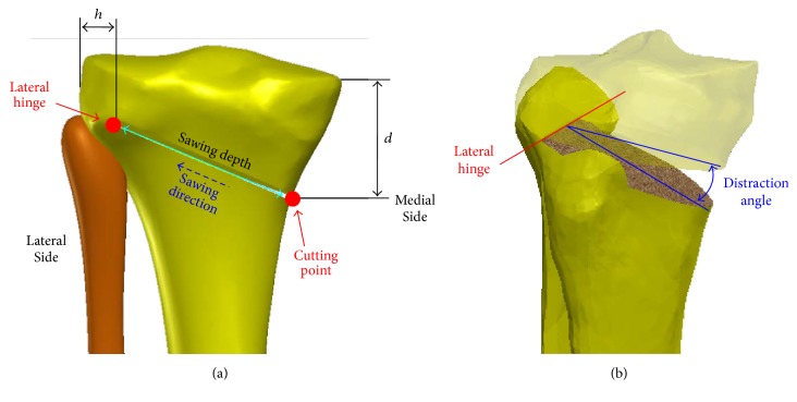
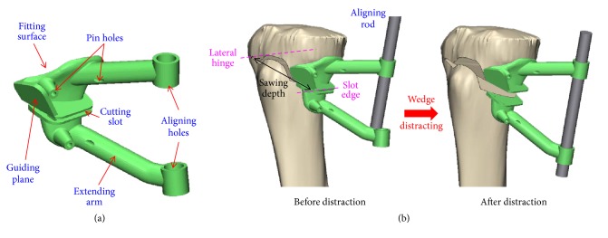
High tibial osteotomy (HTO) has been adopted as an effective surgery for medial degeneration of the osteoarthritis (OA) knee. However, satisfactory outcomes necessitate the precise creation and distraction of osteotomized wedges and the use of intraoperative X-ray images to continually monitor the wedge-related manipulation. Thus HTO is highly technique-demanding and has a high radiation exposure. We report a patient-specific instrument (PSI) guide for the precise creation and distraction of HTO wedge.
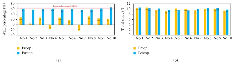
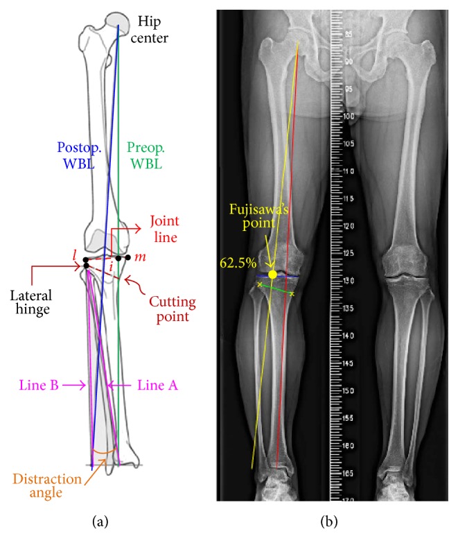
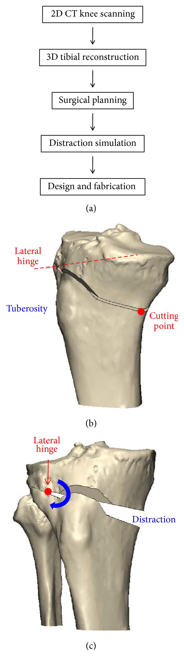
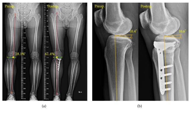
This study first parameterized five HTO procedures to serve as a design rationale for an innovative PSI guide. Preoperative X-ray and computed tomography- (CT-) scanning images were used to design and fabricate PSI guides for clinical use. The weight-bearing line (WBL) of the ten patients was shifted to the Fujisawa's point and instrumented using the TomoFix system. The radiological results of the PSI-guided HTO surgery were evaluated by the WBL percentage and tibial slope.
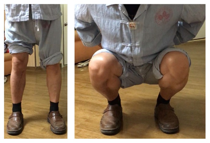
All patients consistently showed an increased range of motion and a decrease in pain and discomfort at about three-month follow-up. This study demonstrates the satisfactory accuracy of the WBL adjustment and tibial slope maintenance after HTO with PSI guide. For all patients, the average pre- and postoperative WBL are, respectively, 14.2% and 60.2%, while the tibial slopes are 9.9 and 10.1 degrees. The standard deviations are 2.78 and 0.36, respectively, in postoperative WBL and tibial slope. The relative errors of the pre- and postoperative WBL percentage and tibial slope averaged 4.9% and 4.1%, respectively.
Instead of using navigator systems, this study integrated 2D and 3D preoperative planning to create a PSI guide that could most likely render the outcomes close to the planning. The PSI guide is a precise procedure that is time-saving, radiation-reducing, and relatively easy to use. Precise osteotomy and good short-term results were achieved with the PSI guide.
胫骨高位截骨术(HTO)已被采纳为治疗膝关节骨关节炎(OA)内侧退变的有效手术。然而,满意的结果需要精确地创建和分离截骨楔形块,并使用术中 X 射线图像来持续监测楔形块相关操作。因此,HTO 技术要求高,辐射暴露量大。我们报告了一种用于精确创建和分离 HTO 楔形块的患者特异性器械(PSI)引导器。
本研究首先对 5 例 HTO 手术进行参数化,作为创新 PSI 引导器的设计原理。使用术前 X 射线和计算机断层扫描(CT)扫描图像来设计和制造用于临床使用的 PSI 引导器。使用 TomoFix 系统将十位患者的负重线(WBL)转移到 Fujisawa 点并进行器械操作。使用 PSI 引导的 HTO 手术后的 WBL 百分比和胫骨斜率评估影像学结果。
所有患者在大约三个月的随访时均表现出运动范围增加,疼痛和不适减轻。本研究表明,PSI 引导的 HTO 手术后 WBL 调整和胫骨斜率维持具有令人满意的准确性。对于所有患者,平均术前和术后 WBL 分别为 14.2%和 60.2%,而胫骨斜率分别为 9.9 和 10.1 度。术后 WBL 和胫骨斜率的标准差分别为 2.78 和 0.36。术前和术后 WBL 百分比和胫骨斜率的相对误差平均分别为 4.9%和 4.1%。
本研究不是使用导航系统,而是整合了 2D 和 3D 术前规划来创建 PSI 引导器,该引导器最有可能使结果接近规划。PSI 引导器是一种精确的手术,节省时间,减少辐射,使用相对简单。PSI 引导器实现了精确的截骨和良好的短期结果。