a Department of Research and Geriatric Research Education and Clinical Center (GRECC) , VA Ann Arbor Healthcare System , Ann Arbor , MI , USA.
b Undergraduate Research Opportunity Program , University of Michigan , Ann Arbor , MI , USA.
J Biomater Sci Polym Ed. 2018 Sep;29(13):1625-1642. doi: 10.1080/09205063.2018.1479084. Epub 2018 Jun 3.
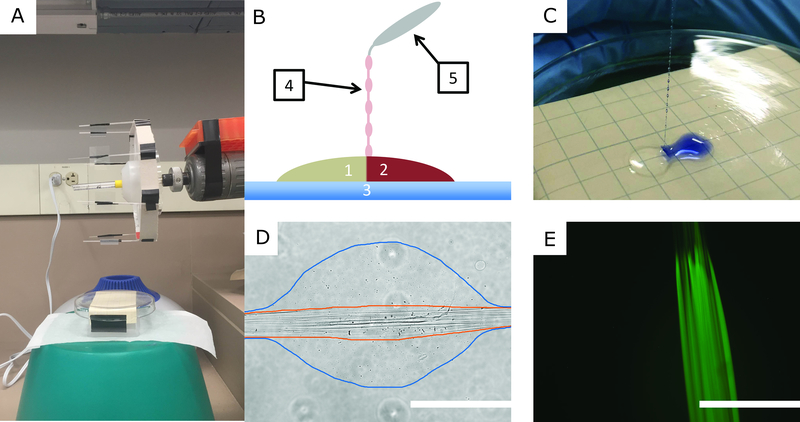
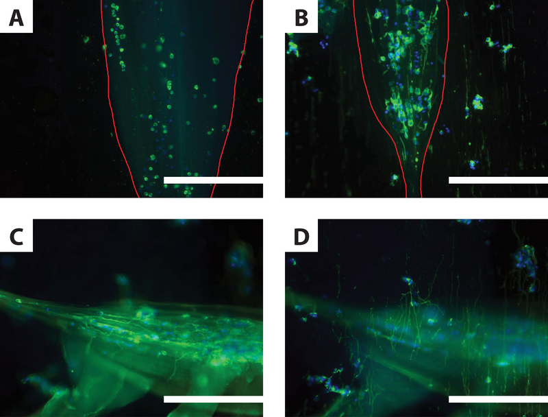
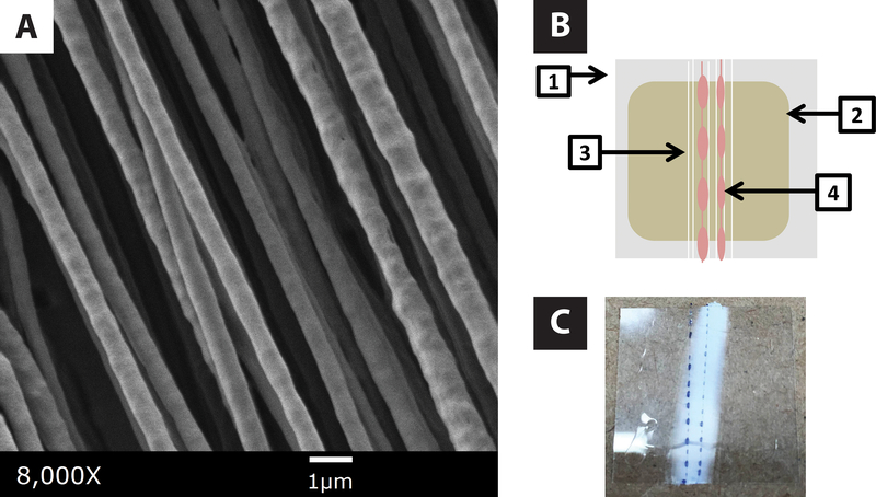
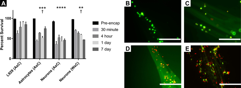
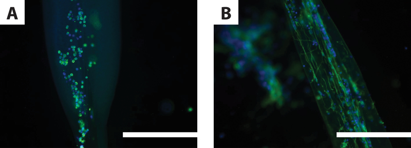
A promising component of biomaterial constructs for neural tissue engineering are electrospun fibers, which differentiate stem cells and neurons as well as direct neurite growth. However, means of protecting neurons, glia, and stem cells seeded on electrospun fibers between lab and surgical suite have yet to be developed. Here we report an effort to accomplish this using cell-encapsulating hydrogel fibers made by interfacial polyelectrolyte complexation (IPC). IPC-hydrogel fibers were created by interfacing acid-soluble chitosan (AsC) and cell-containing alginate and spinning them on bundles of aligned electrospun fibers. Primary spinal astrocytes, cortical neurons, or L929 fibroblasts were mixed into alginate hydrogels prior to IPC-fiber spinning. The viability of each cell type was assessed at 30 min, 4 h, 1 d, and 7 d after encapsulation in IPC hydrogels. Some neurons were encapsulated in IPC-hydrogel fibers made from water-soluble chitosan (WsC). Neurons were also stained with Tuj1 and assessed for neurite extension. Neuron survival in AsC-fibers was worse than astrocytes in AsC-fibers (p < 0.05) and neurons in WsC-fibers (p < 0.05). As expected, neuron and glia survival was worse than L929 fibroblasts (p < 0.05). Neurons in IPC-hydrogel fibers fabricated with WsC extended neurites robustly, while none in AsC fibers did. Neurons remaining inside IPC-hydrogel fibers extended neurites inside them, while others de-encapsulated, extending neurites on electrospun fibers, which did not fully integrate with IPC-hydrogel fibers. This study demonstrates that primary neurons and astrocytes can be encapsulated in IPC-hydrogel fibers at good percentages of survival. IPC hydrogel technology may be a useful tool for encapsulating neural and other cells on electrospun fiber scaffolds.
用于神经组织工程的生物材料构建的有前途的组成部分是电纺纤维,它可以分化干细胞和神经元,并直接引导神经突生长。然而,在实验室和手术室内保护接种在电纺纤维上的神经元、神经胶质细胞和干细胞的方法尚未开发出来。在这里,我们报告了使用界面聚电解质络合(IPC)来实现这一目标的努力。通过界面酸溶性壳聚糖(AsC)和含有细胞的海藻酸钠之间的相互作用,然后将它们纺制到排列整齐的电纺纤维束上,形成 IPC-水凝胶纤维。将原代脊髓星形胶质细胞、皮质神经元或 L929 成纤维细胞混合到藻酸盐水凝胶中,然后进行 IPC 纤维纺丝。在 IPC 水凝胶包封后 30 分钟、4 小时、1 天和 7 天,评估每种细胞类型的活力。将一些神经元包封在由水溶性壳聚糖(WsC)制成的 IPC-水凝胶纤维中。还对神经元进行了 Tuj1 染色并评估了其突起延伸情况。与 AsC 纤维中的星形胶质细胞相比,AsC 纤维中的神经元的存活率更差(p<0.05),与 WsC 纤维中的神经元相比(p<0.05)也是如此。不出所料,神经元和神经胶质细胞的存活率比 L929 成纤维细胞差(p<0.05)。在由 WsC 制成的 IPC 水凝胶纤维中,神经元可以很好地延伸神经突,而在 AsC 纤维中则没有。仍留在 IPC 水凝胶纤维内的神经元在纤维内延伸其神经突,而其他神经元则去包封,在电纺纤维上延伸其神经突,这些纤维没有完全与 IPC 水凝胶纤维整合。这项研究表明,原代神经元和星形胶质细胞可以以较高的存活率被包封在 IPC 水凝胶纤维中。IPC 水凝胶技术可能是一种有用的工具,可用于将神经细胞和其他细胞包封在电纺纤维支架上。