Department of Chemistry and Biochemistry , University of California at San Diego , 9500 Gilman Drive , La Jolla , California 92093 , United States.
Institute of Biomedical Engineering and Informatics , Technische Universität Ilmenau , Gustav-Kirchhoff-Straße , 298693 Ilmenau , Germany.
ACS Appl Mater Interfaces. 2019 Jan 9;11(1):356-372. doi: 10.1021/acsami.8b18344. Epub 2018 Dec 19.
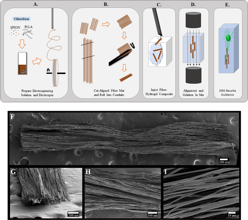
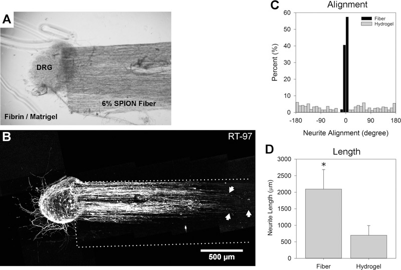
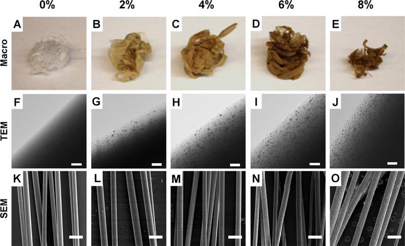
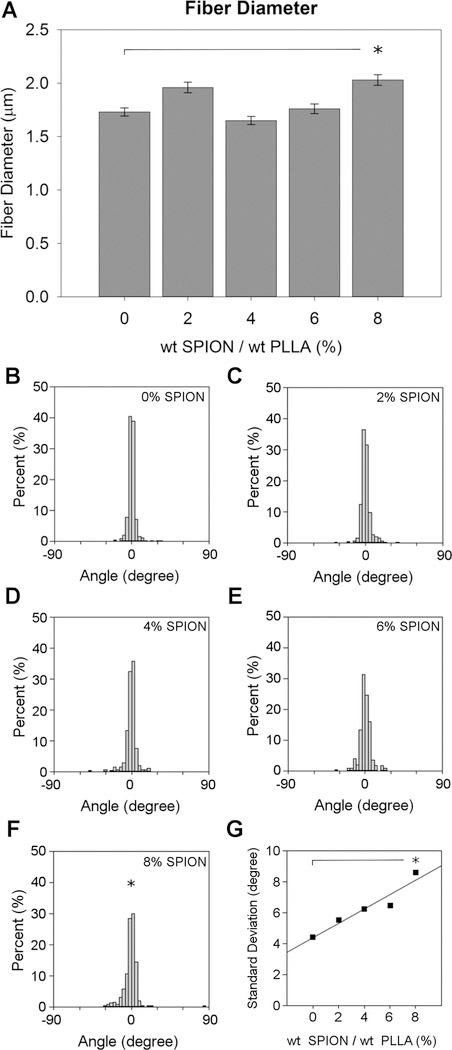
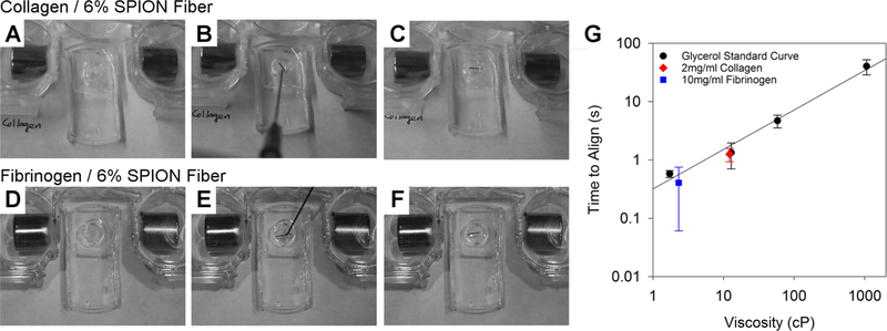
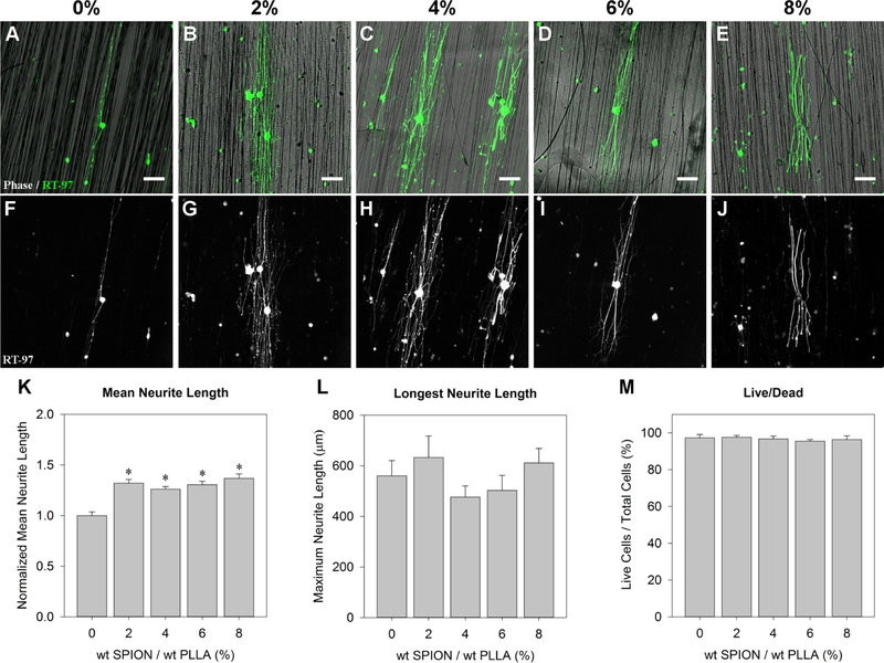
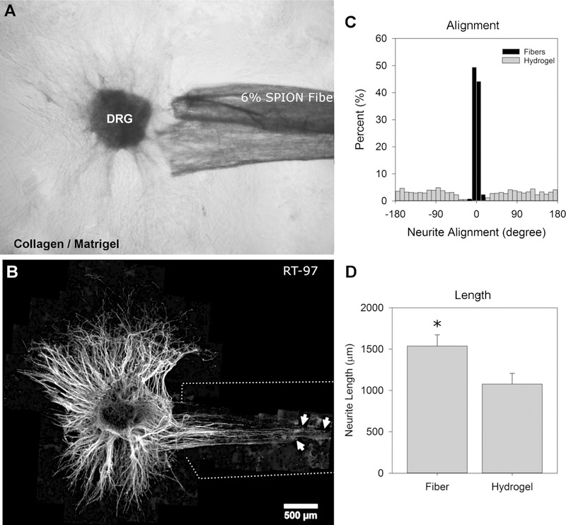
Magnetic electrospun fibers are of interest for minimally invasive biomaterial applications that also strive to provide cell guidance. Magnetic electrospun fibers can be injected and then magnetically positioned in situ, and the aligned fiber scaffolds provide consistent topographical guidance to cells. In this study, magnetically responsive aligned poly-l-lactic acid electrospun fiber scaffolds were developed and tested for neural applications. Incorporating oleic acid-coated iron oxide nanoparticles significantly increased neurite outgrowth, reduced the fiber alignment, and increased the surface nanotopography of the electrospun fibers. After verifying neuron viability on two-dimensional scaffolds, the system was tested as an injectable three-dimensional scaffold. Small conduits of aligned magnetic fibers were easily injected in a collagen or fibrinogen hydrogel solution and repositioned using an external magnetic field. The aligned magnetic fibers provided internal directional guidance to neurites within a three-dimensional collagen or fibrin model hydrogel, supplemented with Matrigel. Neurites growing from dorsal root ganglion explants extended 1.4-3× farther on the aligned fibers compared with neurites extending in the hydrogel alone. Overall, these results show that magnetic electrospun fiber scaffolds can be injected and manipulated with a magnetic field in situ to provide directional guidance to neurons inside an injectable hydrogel. Most importantly, this injectable guidance system increased both neurite alignment and neurite length within the hydrogel scaffold.
磁性静电纺纤维在微创生物材料应用中很有意义,这些应用也努力提供细胞导向。磁性静电纺纤维可以注射,并通过磁场在原位定位,而排列的纤维支架为细胞提供一致的地形导向。在这项研究中,开发了对磁性有响应的排列聚 L-乳酸静电纺纤维支架,并将其用于神经应用进行测试。掺入油酸包覆的氧化铁纳米粒子显著增加了神经突的生长,降低了纤维的排列,并增加了静电纺纤维的表面纳米形貌。在二维支架上验证了神经元活力后,该系统被测试为可注射的三维支架。排列的磁性纤维的小导管很容易在胶原或纤维蛋白原水凝胶溶液中注射,并通过外部磁场重新定位。排列的磁性纤维为三维胶原或纤维蛋白模型水凝胶中的神经突提供了内部定向导向,补充了基质胶。与单独在水凝胶中延伸的神经突相比,从背根神经节外植体生长的神经突在排列的纤维上延伸了 1.4-3 倍。总的来说,这些结果表明,磁性静电纺纤维支架可以注射,并通过磁场在原位操纵,为可注射水凝胶中的神经元提供定向导向。最重要的是,这种可注射的导向系统增加了水凝胶支架内神经突的排列和长度。