Kim Hye Rin, Jung Hae Kyoung
Department of Radiology, CHA Gangnam Medical Center, CHA University, Seoul, Republic of Korea.
Department of Radiology, CHA Bundang Medical Center, CHA University, Seoul, Republic of Korea.
Acta Radiol Open. 2018 May 29;7(6):2058460118774957. doi: 10.1177/2058460118774957. eCollection 2018 Jun.
There is little research done on non-mass cancers (NMCs) on breast ultrasound (US).
To evaluate large-sectional histopathology findings of NMCs on breast US.
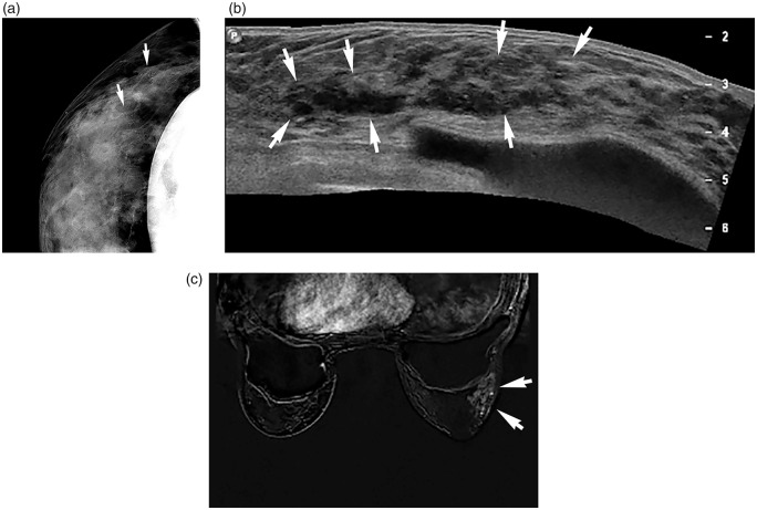
The mammographic and histopathology features of biopsy proven 36 breast cancers which showed pure non-mass lesions on US were retrospectively reviewed.
The most common mammographic finding was microcalcification (23/35, 65.7%); fine pleomorphic microcalcification was predominant (18/23, 78.3%). The main tumor type was pure ductal carcinoma in situ (DCIS) (14/36, 38.9%) and DCIS with micro- or minimal invasion (11/36, 30.6%). Among the 25 DCIS, histologic grade was high in 15 (60.0%) and intermediate in nine (36%); comedo necrosis was seen in 17 (68%). Immunohistochemical analysis was available in 27 lesions and showed HER2-overexpression in 12 (44.4%) and triple-negative in two (7.4%).
According to our limited patient sample, NMCs on breast US were mainly associated with high-grade DCIS.
关于乳腺超声(US)检查中的非肿块性癌(NMCs)的研究较少。
评估乳腺超声检查中NMCs的大体组织病理学表现。
对36例经活检证实的乳腺癌患者的乳腺X线摄影和组织病理学特征进行回顾性分析,这些患者在超声检查中均表现为单纯的非肿块性病变。
乳腺X线摄影最常见的表现为微钙化(23/35,65.7%);细多形性微钙化为主(18/23,78.3%)。主要肿瘤类型为单纯导管原位癌(DCIS)(14/36,38.9%)和伴有微浸润或最小浸润的DCIS(11/36,30.6%)。在25例DCIS中,15例(60.0%)组织学分级为高级别,9例(36%)为中级别;17例(68%)可见粉刺样坏死。27个病灶可进行免疫组化分析,其中12例(44.4%)显示HER2过表达,2例(7.4%)为三阴性。
根据我们有限的患者样本,乳腺超声检查中的NMCs主要与高级别DCIS相关。