Radiological Sciences, Division of Clinical Neuroscience, School of Medicine, University of Nottingham, Queen's Medical Centre, Nottingham, UK; Sir Peter Mansfield Imaging Centre, University of Nottingham, Nottingham, UK; Department of Radiology, Cardiff and Vale University Health Board, Cardiff, UK; Cardiff University Brain Research Imaging Centre (CUBRIC), University of Cardiff, Cardiff, UK.
Sir Peter Mansfield Imaging Centre, University of Nottingham, Nottingham, UK.
Neuroimage Clin. 2018 May 24;19:683-689. doi: 10.1016/j.nicl.2018.05.027. eCollection 2018.
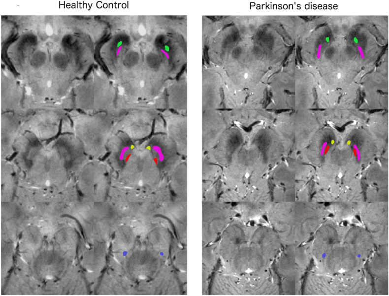
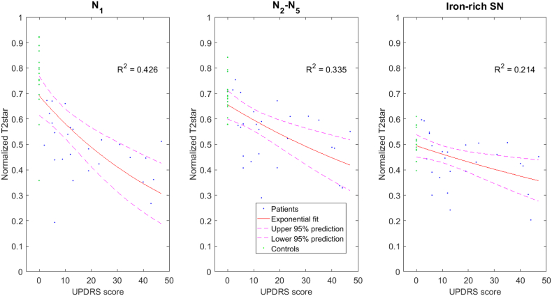
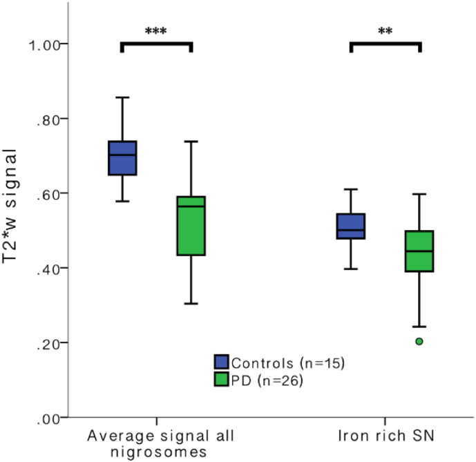
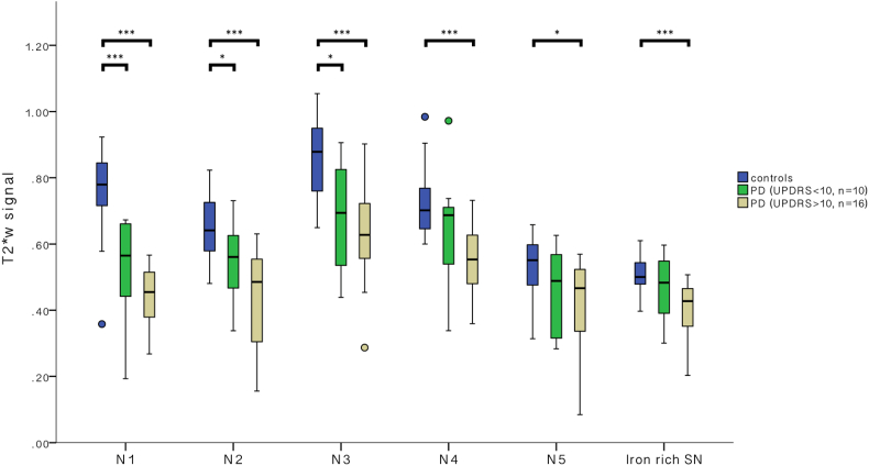
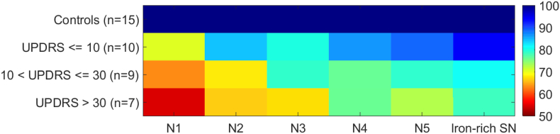
Improved markers for the progression of Parkinson's disease (PD) are required. Previous work has proven that iron dependent MRI scans can detect the largest Nigrosome (N1) within the substantia nigra (SN) pars compacta and changes in PD. Histopathological studies have shown that N1 is particularly affected in early PD whereas the other nigrosomes (N2-N5) and the surrounding iron-rich SN are affected later. In this study we aimed to determine whether MRI can detect the smaller nigrosomes (N2-N5) and whether graded signal alterations can be detected on T2*-weighted MRI at different disease stages consistent with histopathological changes. An observational prospective study was performed within the research imaging centre at the University of Nottingham, UK. Altogether 26 individuals with confirmed PD (median Hoehn&Yahr stage = 1, Unified PD Rating Scale [UPDRS] = 12.5) and 15 healthy controls participated. High resolution T2weighted 7T MRI of the brain was performed and visibility of N1-N5 within the SN was qualitatively rated. Normalised T2weighted signal intensities in manually segmented N1-N5 regions and iron-rich SN were calculated. We performed group comparisons and correlations with severity based on UPDRS. Qualitative measures were a nigrosome visibility score and a confidence score for identification. Quantitative measures were T2weighted contrast of N1-5 and iron-rich SN relative to white matter. We found that visual assessment of the SN for N1-N5 revealed normal range visibility scores in 14 of 15 controls. N1 was identified with the highest confidence and visibility was in abnormal range in all 26 PD patients. The other nigrosomes were less well visible and less confidently identified. There was a larger PD induced signal reduction in all nigrosomes than in the iron-rich SN (median signal difference N1-5 PD compared to controls: 19.4% [IQR = 24%], iron-rich SN 11% [IQR = 24%, = 0.017]). The largest PD induced signal reduction was in N1: 37.2% [IQR = 19%] which inversely correlated with UPDRS in PD (R = 0.19). All nigrosomes can be detected using 7T MRI, and PD induced T2weighted signal reduction was greatest in the nigrosomes (especially N1). The graded T2weighted signal alterations in the nigrosomes match previously described differential histopathological effects of PD. N1 was identified with the highest confidence and T2weighted signal in N1 correlated with UPDRS confirming N1 as the most promising SN marker of PD pathology.
需要更好的帕金森病(PD)进展标志物。先前的工作已经证明,铁依赖性 MRI 扫描可以检测到黑质(SN)致密部中最大的 Nigrosome(N1)和 PD 的变化。组织病理学研究表明,在早期 PD 中,N1 受到的影响特别大,而其他 Nigrosomes(N2-N5)和周围富含铁的 SN 则受到的影响较晚。在这项研究中,我们旨在确定 MRI 是否可以检测到较小的 Nigrosomes(N2-N5),以及在不同疾病阶段是否可以在 T2*-加权 MRI 上检测到分级信号改变,这些改变与组织病理学变化一致。在英国诺丁汉大学的研究成像中心进行了一项观察性前瞻性研究。共有 26 名确诊的 PD 患者(中位 Hoehn&Yahr 分期 = 1,统一 PD 评定量表 [UPDRS] = 12.5)和 15 名健康对照者参与了研究。对大脑进行了高分辨率 T2加权 7T MRI 扫描,并对 SN 内的 N1-N5 进行了定性评分。计算了手动分割的 N1-N5 区域和富含铁的 SN 中的归一化 T2加权信号强度。我们根据 UPDRS 进行了组间比较和严重程度的相关性分析。定性指标是 Nigrosome 可视性评分和识别置信度评分。定量指标是相对于白质的 N1-5 和富含铁的 SN 的 T2加权对比。我们发现,对 SN 中 N1-N5 的视觉评估显示,15 名对照者中有 14 名的可视范围得分正常。在所有 26 名 PD 患者中,N1 的识别率最高,且可视范围异常。其他 Nigrosomes的可视性较差,识别度也较低。与富含铁的 SN 相比,所有 Nigrosomes 的 PD 诱导信号降低幅度更大(N1-5 PD 与对照组相比的中位数信号差异:19.4% [IQR = 24%],富含铁的 SN 为 11% [IQR = 24%,= 0.017])。最大的 PD 诱导信号降低发生在 N1:37.2% [IQR = 19%],这与 PD 中的 UPDRS 呈负相关(R = 0.19)。所有 Nigrosomes 都可以使用 7T MRI 检测到,并且 PD 诱导的 T2加权信号降低在 Nigrosomes 中最大(尤其是 N1)。Nigrosomes 中分级的 T2加权信号改变与先前描述的 PD 的不同组织病理学效应相匹配。N1 的识别置信度最高,N1 中的 T2加权信号与 UPDRS 相关,证实 N1 是 PD 病理最有前途的 SN 标志物。