Fan Qiuyun, Nummenmaa Aapo, Wichtmann Barbara, Witzel Thomas, Mekkaoui Choukri, Schneider Walter, Wald Lawrence L, Huang Susie Y
Athinoula A. Martinos Center for Biomedical Imaging, Department of Radiology, Massachusetts General Hospital, Harvard Medical School, 149 Thirteenth Street, Suite 2301, Charlestown, MA, United States.
Computer Assisted Clinical Medicine, Medical Faculty Mannheim, Heidelberg University, Mannheim, Germany.
Data Brief. 2018 Mar 12;18:334-339. doi: 10.1016/j.dib.2018.03.021. eCollection 2018 Jun.
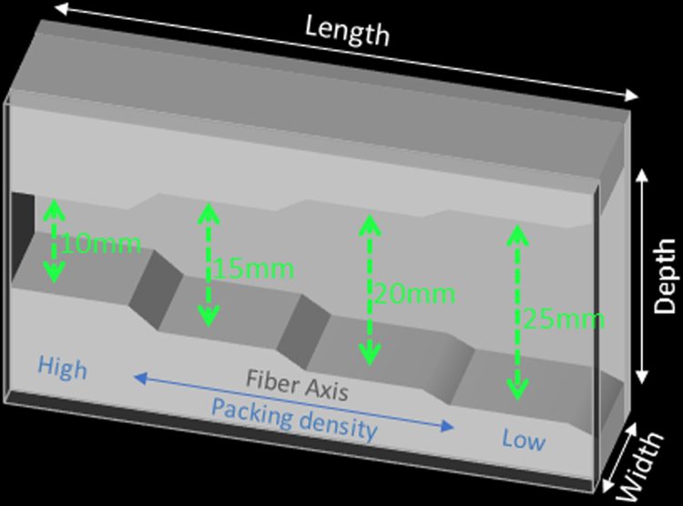
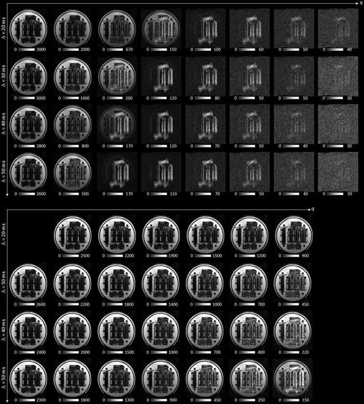
We provide a comprehensive diffusion MRI dataset acquired with a novel biomimetic phantom mimicking human white matter. The fiber substrates in the diffusion phantom were constructed from hollow textile axons ("taxons") with an inner diameter of 11.8±1.2 µm and outer diameter of 33.5±2.3 µm. Data were acquired on the 3 T CONNECTOM MRI scanner with multiple diffusion times and multiple q-values per diffusion time, which is a dedicated acquisition for validation of microstructural imaging methods, such as compartment size and volume fraction mapping. Minimal preprocessing was performed to correct for susceptibility and eddy current distortions. Data were deposited in the XNAT Central database (project ID: dMRI_Phant_MGH).
我们提供了一个通过模仿人类白质的新型仿生体模采集的综合扩散磁共振成像(dMRI)数据集。扩散体模中的纤维基质由内径为11.8±1.2微米、外径为33.5±2.3微米的空心纺织轴突(“分类单位”)构建而成。数据在3T CONNECTOM磁共振成像扫描仪上采集,每个扩散时间有多个扩散时间点和多个q值,这是用于验证微观结构成像方法(如隔室大小和体积分数映射)的专用采集方式。进行了最少的预处理以校正磁化率和涡流畸变。数据已存入XNAT中央数据库(项目ID:dMRI_Phant_MGH)。