Shanghai Key Laboratory of Forensic Medicine, Shanghai Forensic Service Platform, Institute of Forensic Science, Ministry of Justice, Shanghai, China (mainland).
Department of Psychology, Qiqihar Mental Health Center, Qiqihar, Heilongjiang, China (mainland).
Med Sci Monit. 2018 Jun 13;24:4020-4030. doi: 10.12659/MSM.905354.
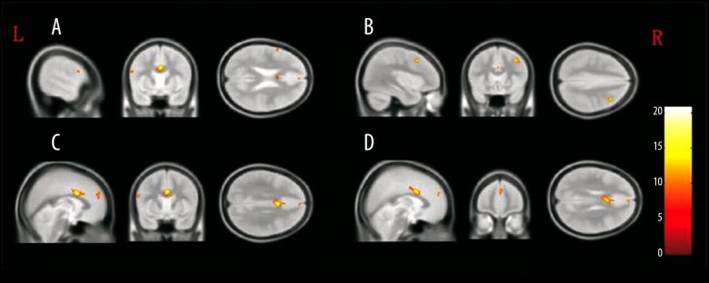
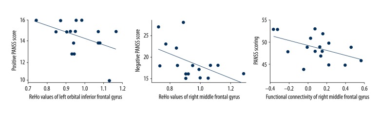
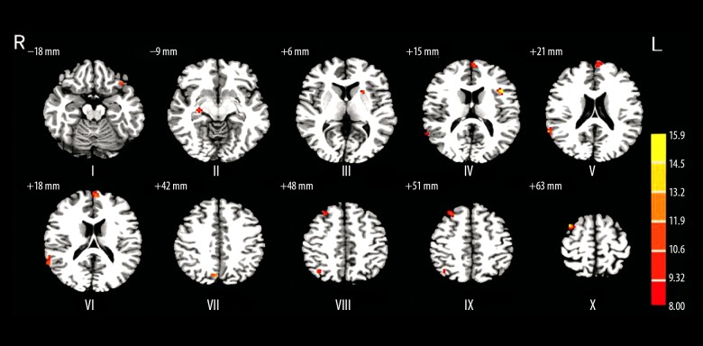
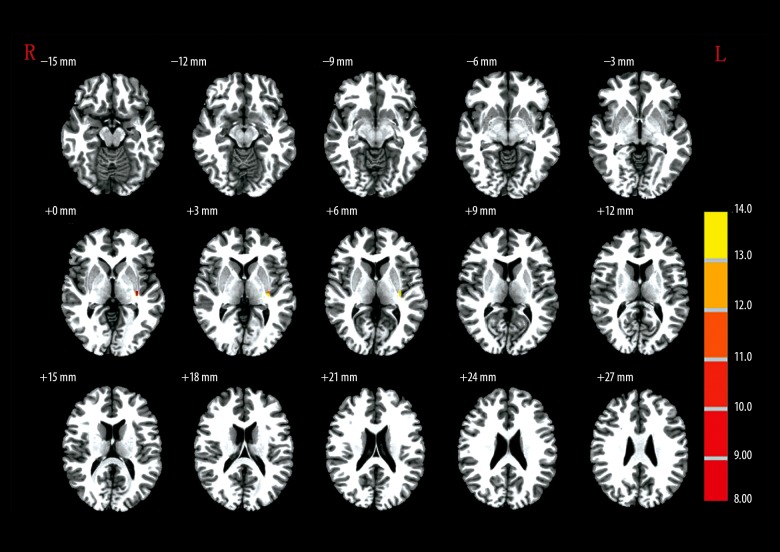
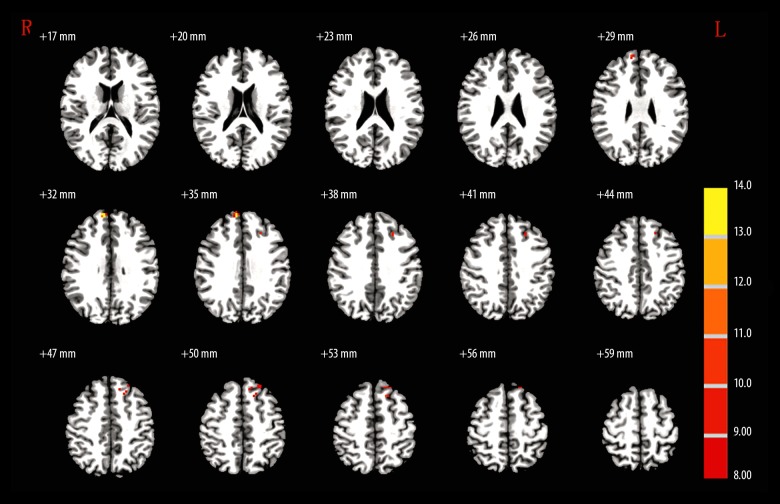
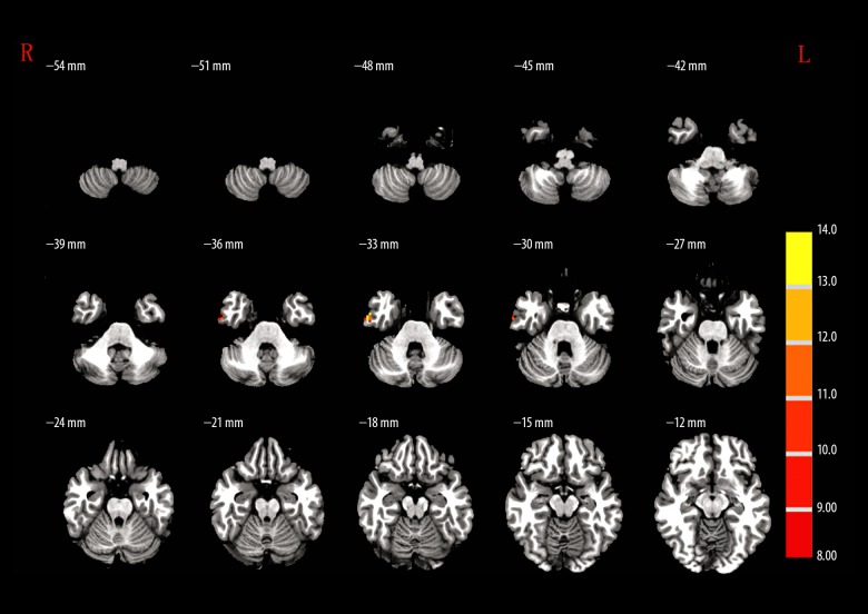
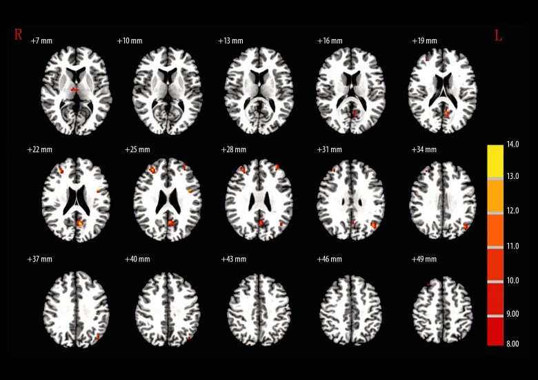
BACKGROUND Using regional homogeneity (ReHo) blood oxygen level-dependent functional MR (BOLD-fMRI), we investigated the structural and functional alterations of brain regions among patients with methamphetamine-associated psychosis (MAP). MATERIAL AND METHODS This retrospective study included 17 MAP patients, 16 schizophrenia (SCZ) patients, and 18 healthy controls. Informed consent was obtained from all patients before the clinical assessment, the severity of clinical symptoms was evaluated prior to the fMRI scanning, and then images were acquired and preprocessed after each participant received 6-min fRMI scanning. The participants all underwent BOLD-fMRI scanning. Voxel-based morphometry was used to measure gray matter density (GMD). Resting-state fMRI (rs-fMRI) was conducted to analyze functional MR, ReHo, and functional connectivity (FC). RESULTS GMD analysis results suggest that MAP patients, SCZ patients, and healthy volunteers show different GMDs within different brain regions. Similarly, the ReHo analysis results suggest that MAP patients, SCZ patients, and healthy volunteers have different GMDs within different brain regions. Negative correlations were found between ReHo- and the PANSS-positive scores within the left orbital interior frontal gyrus (L-orb-IFG) of MAP patients. ReHo- and PANSS-negative scores of R-SFG were negatively correlated among SCZ patients. The abnormal FC of R-MFG showed a negative correlation with the PANSS score among MAP patients. CONCLUSIONS The abnormalities in brain structure and FC were associated with the development of MAP.
使用局部一致性(ReHo)血氧水平依赖功能磁共振(BOLD-fMRI),我们研究了与甲基苯丙胺相关精神病(MAP)患者的脑区结构和功能改变。
这是一项回顾性研究,纳入了 17 例 MAP 患者、16 例精神分裂症(SCZ)患者和 18 例健康对照者。所有患者在临床评估前均获得知情同意,在 fMRI 扫描前评估了临床症状的严重程度,然后在每位参与者接受 6 分钟 fMRI 扫描后采集和预处理图像。所有参与者均接受 BOLD-fMRI 扫描。基于体素的形态计量学用于测量灰质密度(GMD)。静息态 fMRI(rs-fMRI)用于分析功能磁共振、ReHo 和功能连接(FC)。
GMD 分析结果表明,MAP 患者、SCZ 患者和健康志愿者在不同脑区显示出不同的 GMD。同样,ReHo 分析结果表明,MAP 患者、SCZ 患者和健康志愿者在不同脑区显示出不同的 GMD。MAP 患者左眶额内侧回(L-orb-IFG)的 ReHo 与 PANSS 阳性评分呈负相关。SCZ 患者的 R-SFG 的 ReHo 与 PANSS 阴性评分呈负相关。MAP 患者的 R-MFG 的异常 FC 与 PANSS 评分呈负相关。
脑结构和 FC 的异常与 MAP 的发生有关。