Zheng Chunhui, Chen Xiaomei, Zhang Fangbiao, Yan Liping, Zhang Xiangyan
Department of Cardiothoracic Surgery Operating Room Department of Pathology, Lishui Hospital of Zhejiang University, Lishui Central Hospital, Lishui, Zhejiang Province, P.R. China.
Medicine (Baltimore). 2018 Jun;97(24):e11165. doi: 10.1097/MD.0000000000011165.
Primary mucoepidermoid carcinoma (MEC) of the esophagus is a rare type of malignant neoplasm. Its morphology resembles that of MEC of the salivary glands. It is characterized by a diffuse mixture of squamous and mucus-secreting glandular carcinoma cells. Due to the low incidence of esophageal MEC, the biological behavior and treatment of this tumor have not been well studied.
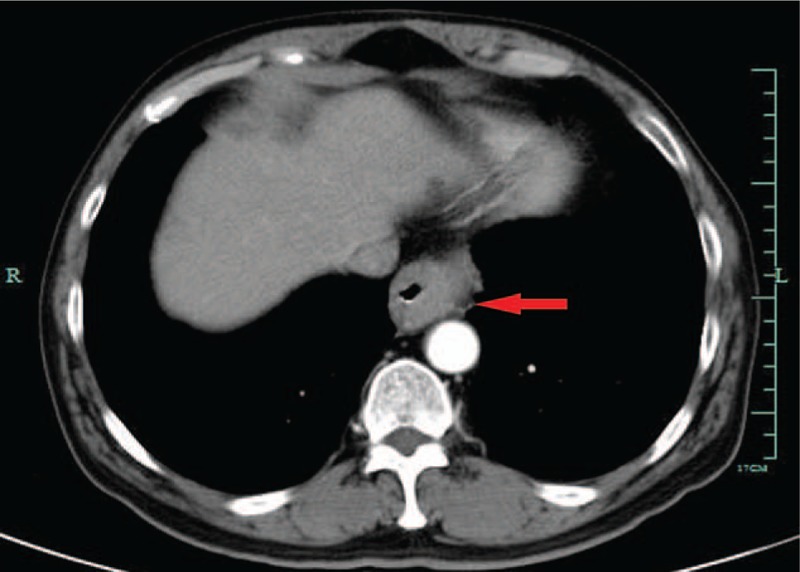
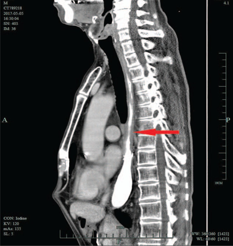
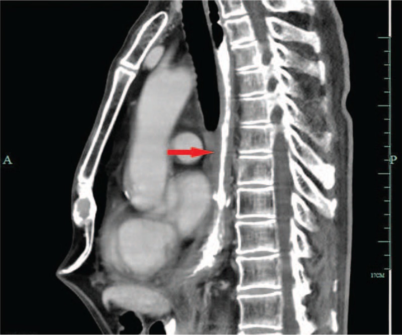
In this case report, we describe a case of a 59-year-old man who presented with difficulty in swallowing. Iohexol swallowing revealed a malignant-appearing structure in the inferior-thoracic region.
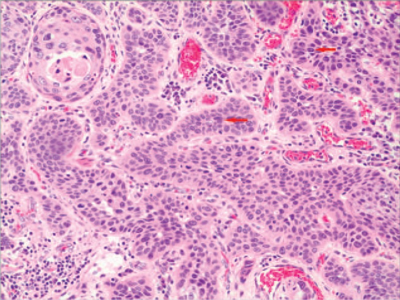
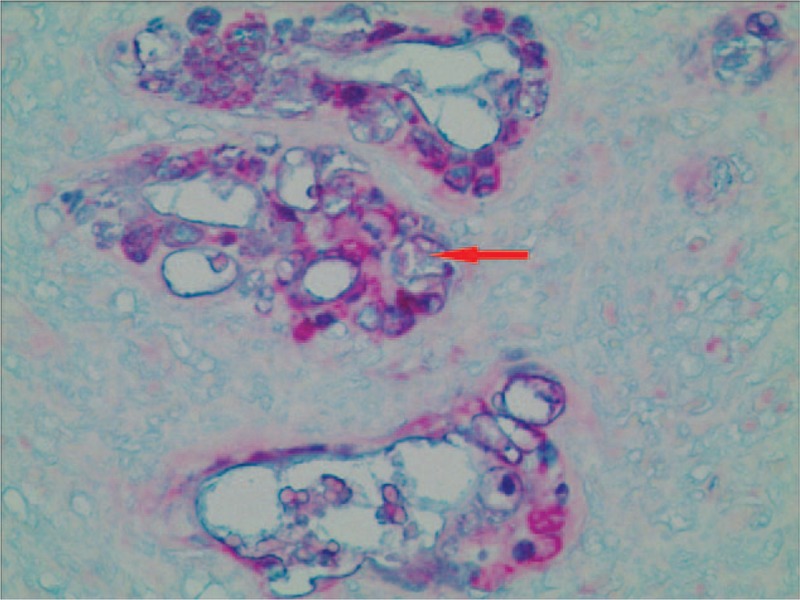
Biopsy of the lesion under endoscopy demonstrated a mucoepidermoid carcinoma of the esophagus.
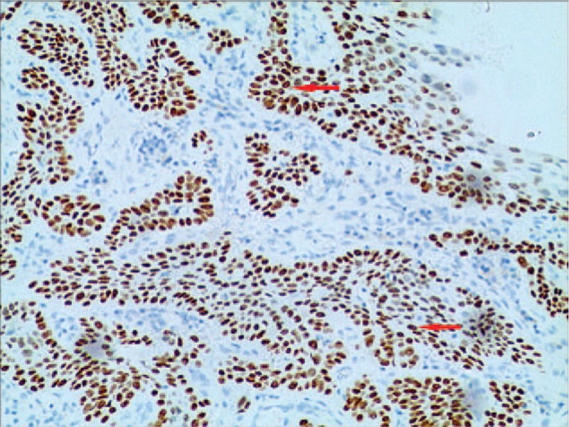
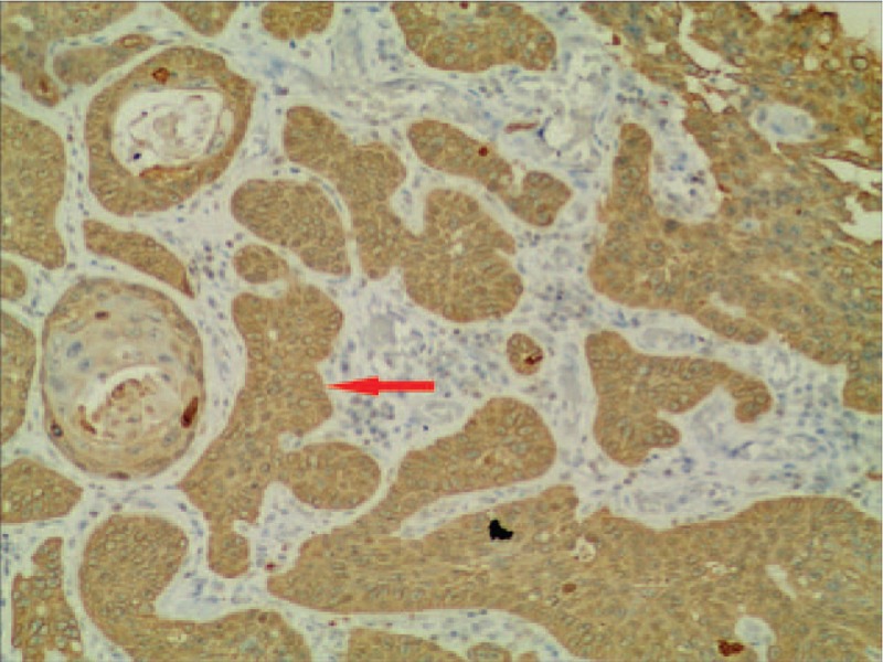
We performed esophagectomy, esophagogastrostomy through the esophageal bed and 2-field lymphadenectomy. Histopathological analysis of the tumor revealed histological characteristics typical of an esophageal MEC. Radio-chemotherapy was administered to this patient.
Seventeen months after surgery, an esophageal computed tomography (CT) scan revealed that the wall of esophagus was evenly thickened. However, endoscopic assessment revealed no evidence of recurrence. Further CT scans at 19 and 31 months after surgery also showed a thickened esophageal wall, although endoscopic assessment at 31 months still revealed no esophageal stricture and no evidence of recurrence. The patient is alive with no dysphagia and no evidence of recurrence for over 39 months.
There is little evidence of effective treatment nor guidelines for treatment of esophageal MEC. Although the general prognosis of esophageal MEC is poor, comprehensive treatment of surgery and radio-chemotherapy appeared to be effective in this case. Radio-chemotherapy is a possible treatment option that was shown to have acceptable short-term effects.
原发性食管黏液表皮样癌(MEC)是一种罕见的恶性肿瘤。其形态与涎腺MEC相似。其特征是鳞状细胞癌和黏液分泌性腺癌细胞的弥漫性混合。由于食管MEC发病率低,该肿瘤的生物学行为和治疗尚未得到充分研究。
在本病例报告中,我们描述了一名59岁男性患者,他出现吞咽困难。碘海醇吞咽检查显示胸段下部有一个疑似恶性的结构。
内镜下病变活检显示为食管黏液表皮样癌。
我们进行了食管切除术、经食管床食管胃吻合术和二野淋巴结清扫术。肿瘤的组织病理学分析显示出食管MEC典型的组织学特征。对该患者进行了放化疗。
术后17个月,食管计算机断层扫描(CT)显示食管壁均匀增厚。然而,内镜评估未发现复发迹象。术后19个月和31个月的进一步CT扫描也显示食管壁增厚,尽管31个月时的内镜评估仍未发现食管狭窄和复发迹象。患者存活,无吞咽困难,超过39个月无复发迹象。
几乎没有证据表明食管MEC有有效的治疗方法,也没有治疗指南。尽管食管MEC的总体预后较差,但在本病例中,手术和放化疗的综合治疗似乎有效。放化疗是一种可能的治疗选择,已显示出可接受的短期效果。