Universidad de Buenos Aires, Facultad de Medicina, Dpto. de Biología Celular, Histología, Embriología y Genética, Ciudad Autónoma de Buenos Aires, Argentina.
CONICET-Universidad de Buenos Aires. Instituto de Biología Celular y Neurociencia "Prof. E. De Robertis¨ (IBCN), Ciudad Autónoma de Buenos Aires, Argentina.
PLoS One. 2018 Jun 18;13(6):e0198838. doi: 10.1371/journal.pone.0198838. eCollection 2018.
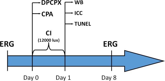
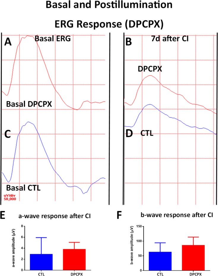
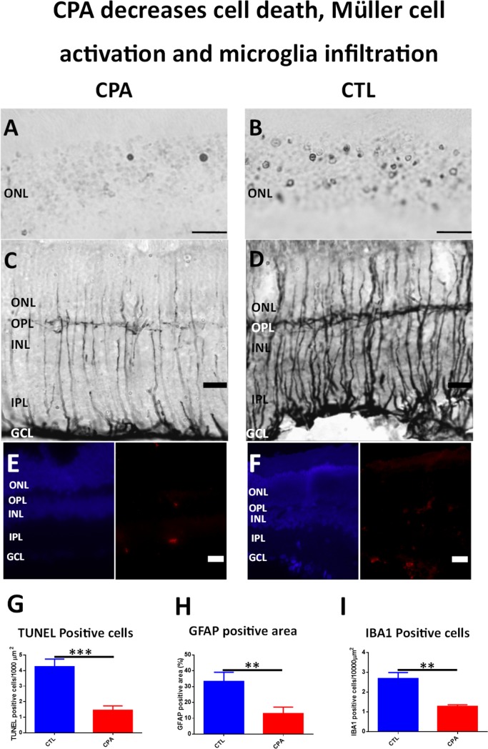
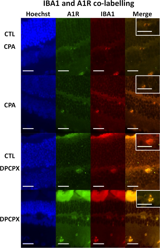
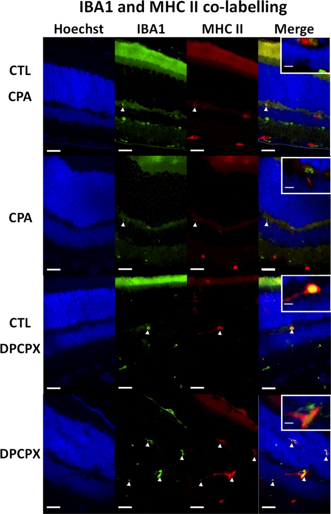
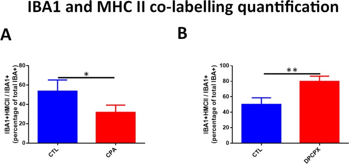
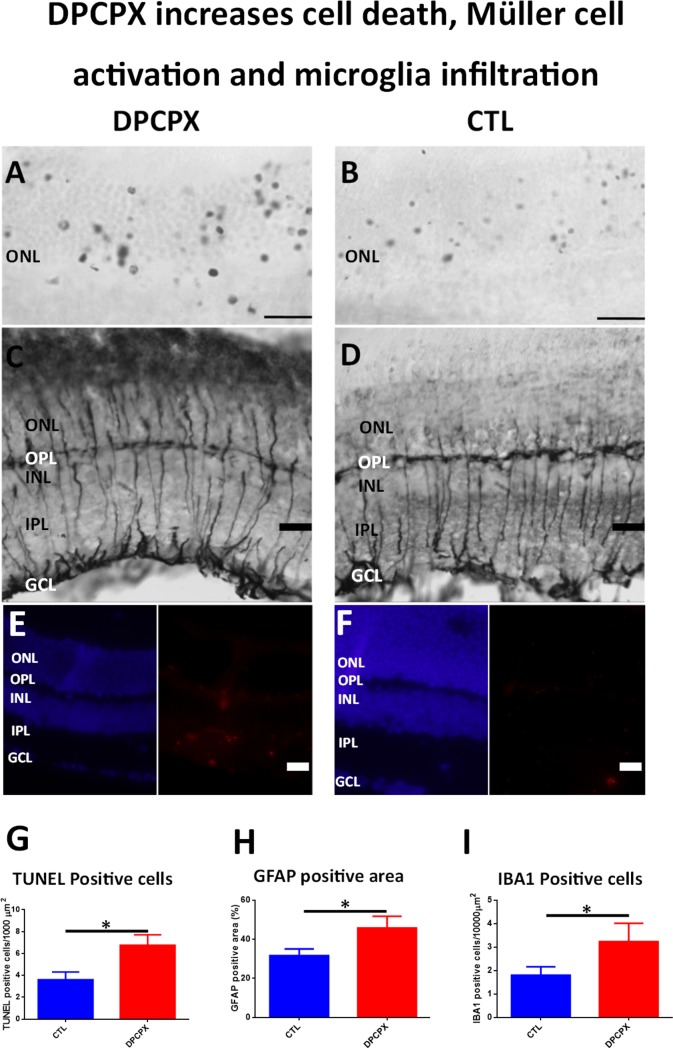
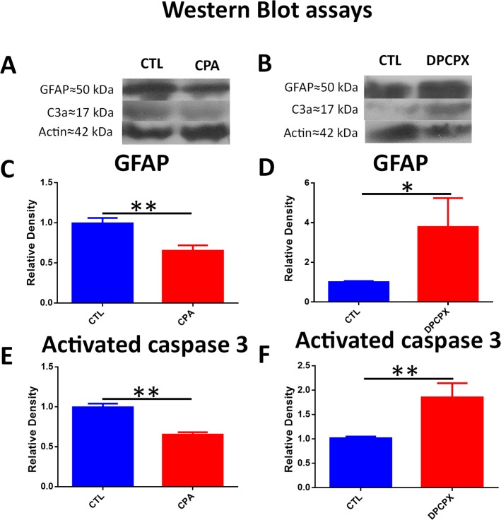
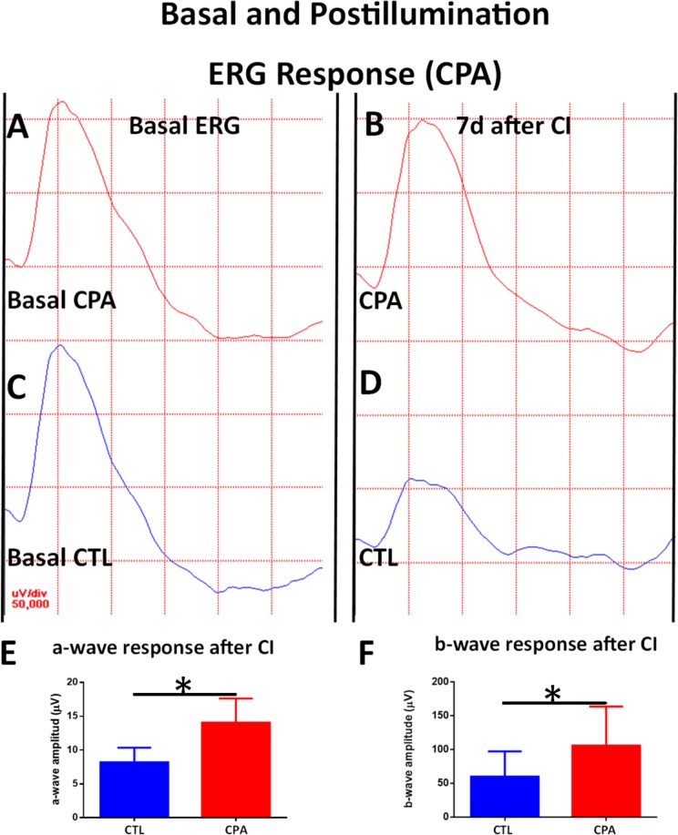
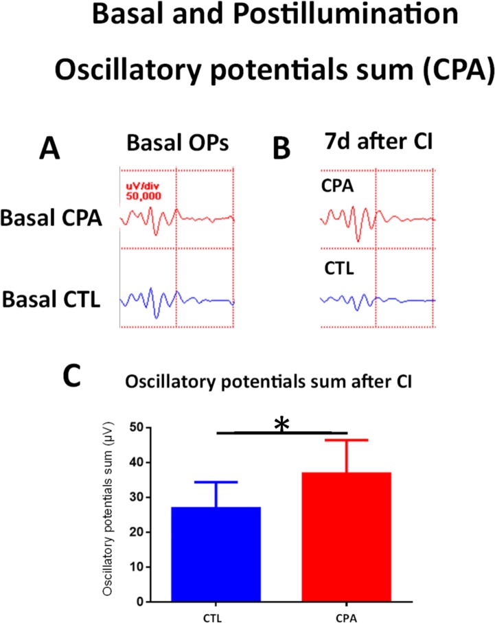
Light induced retinal degeneration (LIRD) is a useful model that resembles human retinal degenerative diseases. The modulation of adenosine A1 receptor is neuroprotective in different models of retinal injury. The aim of this work was to evaluate the potential neuroprotective effect of the modulation of A1 receptor in LIRD. The eyes of rats intravitreally injected with N6-cyclopentyladenosine (CPA), an A1 agonist, which were later subjected to continuous illumination (CI) for 24 h, showed retinas with a lower number of apoptotic nuclei and a decrease of Glial Fibrillary Acidic Protein (GFAP) immunoreactive area than controls. Lower levels of activated Caspase 3 and GFAP were demonstrated by Western Blot (WB) in treated animals. Also a decrease of iNOS, TNFα and GFAP mRNA was demonstrated by RT-PCR. A decrease of Iba 1+/MHC-II+ reactive microglial cells was shown by immunohistochemistry. Electroretinograms (ERG) showed higher amplitudes of a-wave, b-wave and oscillatory potentials after CI compared to controls. Conversely, the eyes of rats intravitreally injected with dipropylcyclopentylxanthine (DPCPX), an A1 antagonist, and subjected to CI for 24 h, showed retinas with a higher number of apoptotic nuclei and an increase of GFAP immunoreactive area compared to controls. Also, higher levels of activated Caspase 3 and GFAP were demonstrated by Western Blot. The mRNA levels of iNOS, nNOS and inflammatory cytokines (IL-1β and TNFα) were not modified by DPCPX treatment. An increase of Iba 1+/MHC-II+ reactive microglial cells was shown by immunohistochemistry. ERG showed that the amplitudes of a-wave, b-wave, and oscillatory potentials after CI were similar to control values. A single pharmacological intervention prior illumination stress was able to swing retinal fate in opposite directions: CPA was neuroprotective, while DPCPX worsened retinal damage. In summary, A1 receptor agonism is a plausible neuroprotective strategy in LIRD.
光诱导视网膜变性(LIRD)是一种有用的模型,类似于人类视网膜退行性疾病。腺苷 A1 受体的调制在不同的视网膜损伤模型中具有神经保护作用。本工作旨在评估调制 A1 受体在 LIRD 中的潜在神经保护作用。向大鼠眼内注射 N6-环戊基腺苷(CPA),一种 A1 激动剂,随后进行 24 小时连续光照(CI),结果显示,与对照组相比,凋亡核数量减少,胶质纤维酸性蛋白(GFAP)免疫反应面积减少。用 Western Blot(WB)证实,用药物处理的动物中的激活 Caspase 3 和 GFAP 水平降低。还通过 RT-PCR 证明 iNOS、TNFα 和 GFAP mRNA 水平降低。免疫组织化学显示 Iba 1+/MHC-II+反应性小胶质细胞减少。视网膜电图(ERG)显示,与对照组相比,CI 后 a 波、b 波和振荡电位的振幅更高。相反,向大鼠眼内注射二丙基环戊基黄嘌呤(DPCPX),一种 A1 拮抗剂,并进行 24 小时 CI,结果显示与对照组相比,凋亡核数量增加,GFAP 免疫反应面积增加。Western Blot 也显示激活 Caspase 3 和 GFAP 的水平升高。DPCPX 处理并未改变 iNOS、nNOS 和炎症细胞因子(IL-1β 和 TNFα)的 mRNA 水平。免疫组织化学显示 Iba 1+/MHC-II+反应性小胶质细胞增加。ERG 显示,CI 后 a 波、b 波和振荡电位的振幅与对照值相似。单次光前应激药物干预能够使视网膜命运向相反方向摆动:CPA 具有神经保护作用,而 DPCPX 则加重视网膜损伤。总之,A1 受体激动剂是 LIRD 中一种合理的神经保护策略。