Center for Life Sciences, Tsinghua University, Beijing, 100084, China; Academy for Advanced Interdisciplinary Studies, Peking University, 100871, Beijing, China.
School of Life Sciences, Peking University, Beijing, 100871, China.
Mol Metab. 2018 Aug;14:71-81. doi: 10.1016/j.molmet.2018.06.004. Epub 2018 Jun 7.
The vascular system is central to sustaining tissue survival and homeostasis. Blood vessels are densely present in adipose tissues and exert essential roles in their metabolism. However, conventional immunohistochemistry methods have intrinsic limitations in examining the 3D vascular network in adipose tissues as well as other organs in general.
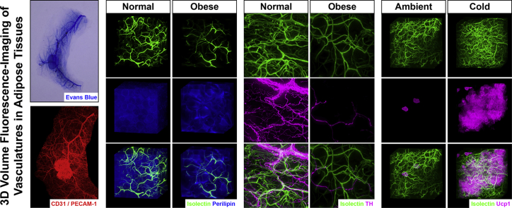
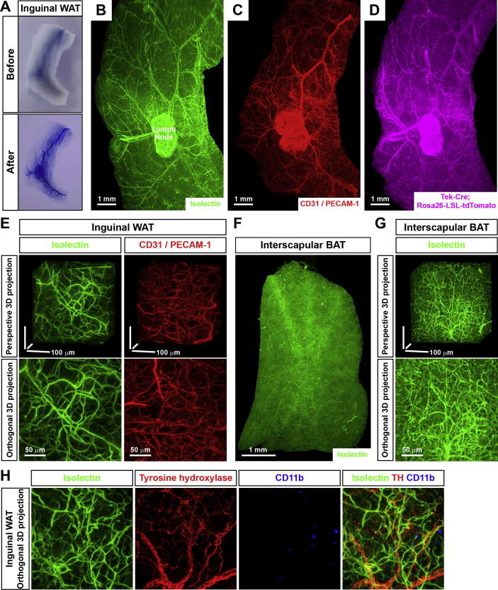
We established a 3D volume fluorescence-imaging technique to visualize the vasculatures in mouse adipose tissues by combining the optimized steps of whole-mount immunolabeling, tissue optical clearing, and lightsheet volume fluorescence-imaging. To demonstrate the strength of this novel imaging procedure, we comprehensively assessed the intra-adipose vasculatures under obese conditions or in response to a cold challenge.
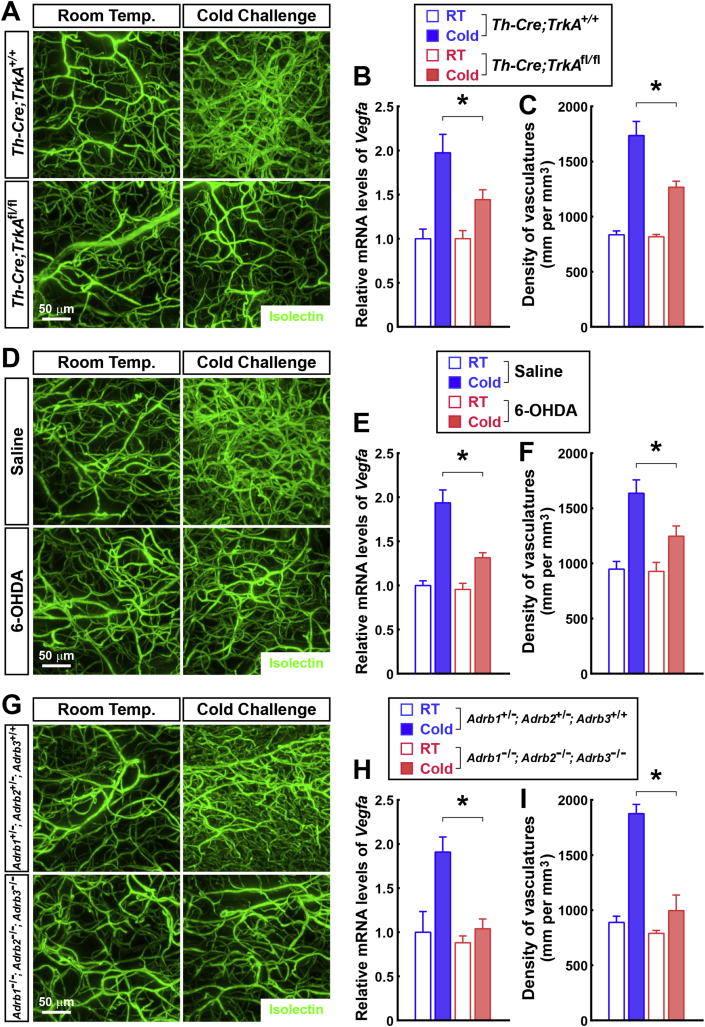
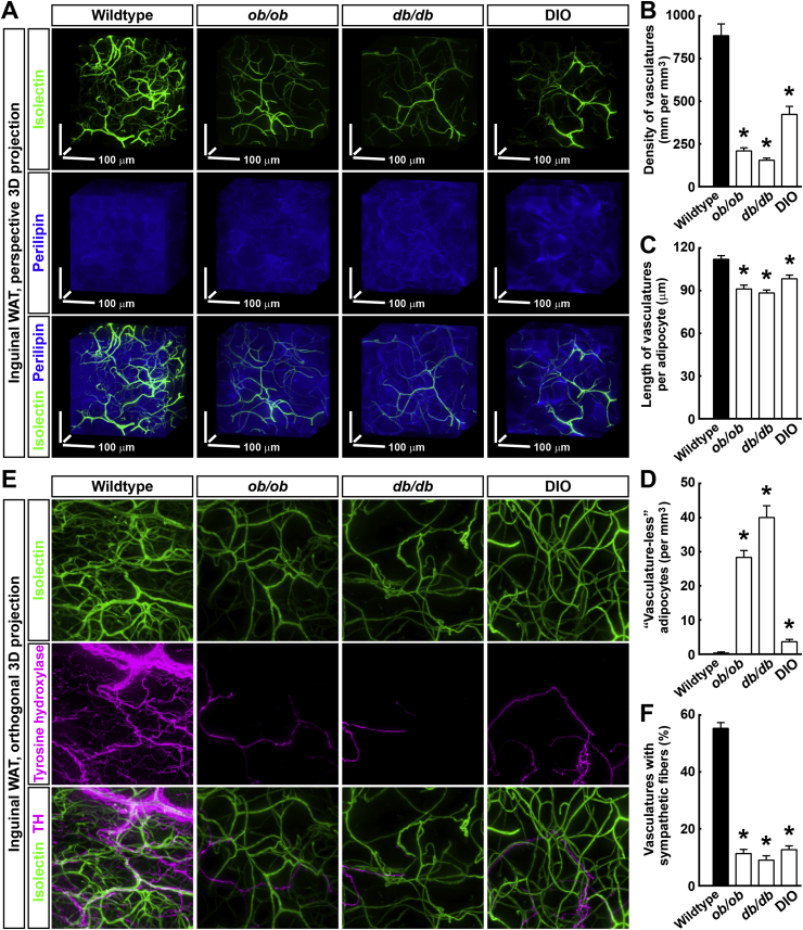
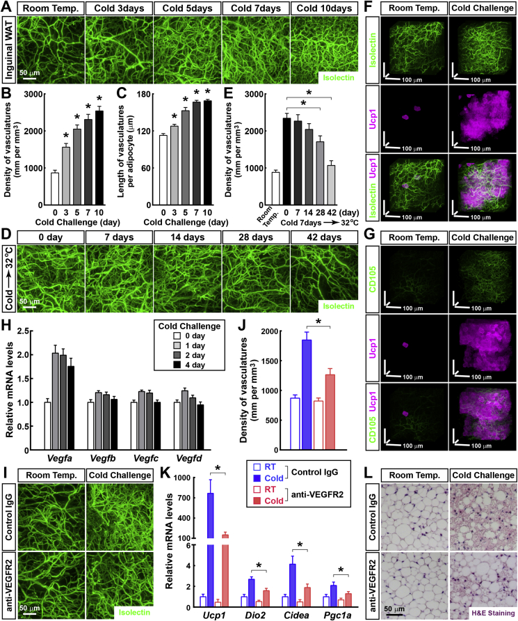
We show the entirety of the vascular network in mouse adipose tissues on the whole-tissue level at a single-capillary resolution for the first time in the field. We accurately quantify the pathological changes of vasculatures in adipose tissues in wild-type or obese mice (ob/ob, db/db, or diet-induced obesity). In addition, we identify significant and reversible changes of the intra-adipose vasculatures in the mice subjected to cold challenge (i.e., 4°). Furthermore, we demonstrate that the cold-induced vascular plasticity depends on the sympathetic-derived catecholamine signal and is involved in the beiging process of white adipose tissues.
We report a 3D volume fluorescence-imaging procedure that is compatible with many areas of vascular research and is poised to serve the field in future investigations of the vascular system in adipose tissues or other research scenarios.
血管系统是维持组织存活和稳态的核心。血管在脂肪组织中密集存在,并在其代谢中发挥重要作用。然而,传统的免疫组织化学方法在检查脂肪组织以及其他器官中的 3D 血管网络方面存在固有局限性。
我们建立了一种 3D 体积荧光成像技术,通过结合全组织免疫标记、组织光学透明化和光片体积荧光成像的优化步骤,可视化小鼠脂肪组织中的脉管系统。为了证明这种新成像程序的优势,我们全面评估了肥胖条件下或冷刺激反应中脂肪组织内的脉管系统。
我们首次在该领域以单毛细血管分辨率在全组织水平上显示了小鼠脂肪组织中的血管网络全貌。我们准确地量化了野生型或肥胖小鼠(ob/ob、db/db 或饮食诱导肥胖)脂肪组织中脉管系统的病理变化。此外,我们发现冷刺激(即 4°C)会导致脂肪组织内脉管系统发生显著且可逆的变化。此外,我们证明了冷诱导的血管可塑性依赖于交感神经源性儿茶酚胺信号,并参与了白色脂肪组织的米色化过程。
我们报告了一种 3D 体积荧光成像程序,该程序与血管研究的许多领域兼容,并有望在未来对脂肪组织或其他研究场景中的血管系统进行研究中为该领域提供服务。