• 文献检索
  • 文档翻译
  • 深度研究
  • 学术资讯
  • Suppr Zotero 插件Zotero 插件
  • 邀请有礼
  • 套餐&价格
  • 历史记录
应用&插件
Suppr Zotero 插件Zotero 插件浏览器插件Mac 客户端Windows 客户端微信小程序
定价
高级版会员购买积分包购买API积分包
服务
文献检索文档翻译深度研究API 文档MCP 服务
关于我们
关于 Suppr公司介绍联系我们用户协议隐私条款
关注我们

Suppr 超能文献

核心技术专利:CN118964589B侵权必究
粤ICP备2023148730 号-1Suppr @ 2026

文献检索

告别复杂PubMed语法,用中文像聊天一样搜索,搜遍4000万医学文献。AI智能推荐,让科研检索更轻松。

立即免费搜索

文件翻译

保留排版,准确专业,支持PDF/Word/PPT等文件格式,支持 12+语言互译。

免费翻译文档

深度研究

AI帮你快速写综述,25分钟生成高质量综述,智能提取关键信息,辅助科研写作。

立即免费体验

高分辨率锥形束CT用于评估颅内部分血栓形成动脉瘤的血管通道。

High-resolution cone beam CT for evaluation of vascular channel in intracranial partial thrombosed aneurysm.

作者信息

Shintai Kazunori, Matsubara Noriaki, Izumi Takashi

机构信息

Department of Neurosurgery, Nagoya University Graduate School of Medicine, Nagoya, Japan.

Department of Neurosurgery, Nagoya Daini Red Cross Hospital, Nagoya, Japan.

出版信息

Nagoya J Med Sci. 2018 May;80(2):279-284. doi: 10.18999/nagjms.80.2.279.

DOI:10.18999/nagjms.80.2.279
PMID:29915445
原文链接:https://pmc.ncbi.nlm.nih.gov/articles/PMC5995734/
Abstract

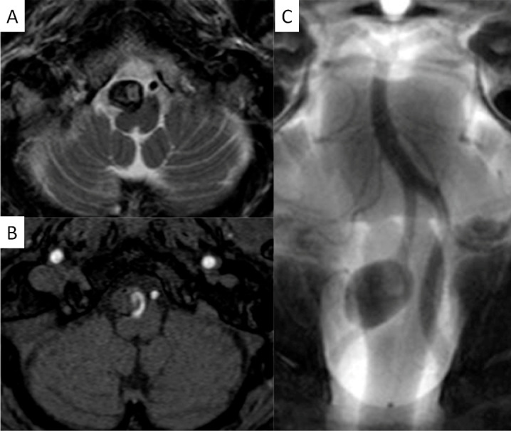
The authors present a 60-year-old man with a partially thrombosed, intracranial vertebral artery aneurysm. A vascular channel in intra-aneurysmal thrombus was effectively identified with high-resolution cone beam CT (DynaCT Micro: Siemens Medical Solutions, Erlangen, Germany). Pre-procedural vertebral angiogram implied a perforating artery arising from near neck of the aneurysm and DynaCT Micro performed before approaching to the lesion demonstrated a vascular channel running in intra-aneurysmal thrombus which could not be distinguished from perforators with other imaging modalities. It was confirmed that perforators around the aneurysm were not identified and safely treated the aneurysm with stent-assisted coil embolization. High-resolution cone beam CT is enable to sharply visualize vessel lumens, thrombus, and intra-thrombus structures, and is useful to identify a vascular channel in intracranial partially thrombosed aneurysm.

摘要

作者报告了一名60岁男性,患有部分血栓形成的颅内椎动脉动脉瘤。通过高分辨率锥形束CT(DynaCT Micro:西门子医疗解决方案公司,德国埃尔朗根)有效地识别了动脉瘤内血栓中的血管通道。术前椎动脉血管造影显示有一条穿支动脉起源于动脉瘤颈部附近,在接近病变前进行的DynaCT Micro显示在动脉瘤内血栓中有一条血管通道,这是其他成像方式无法与穿支动脉区分开来的。证实未识别出动脉瘤周围的穿支动脉,并采用支架辅助弹簧圈栓塞术安全地治疗了该动脉瘤。高分辨率锥形束CT能够清晰地显示血管腔、血栓和血栓内结构,有助于识别颅内部分血栓形成的动脉瘤中的血管通道。

https://cdn.ncbi.nlm.nih.gov/pmc/blobs/5db6/5995734/20d20693d79e/2186-3326-80-0279-g004.jpg
https://cdn.ncbi.nlm.nih.gov/pmc/blobs/5db6/5995734/872de70117e5/2186-3326-80-0279-g001.jpg
https://cdn.ncbi.nlm.nih.gov/pmc/blobs/5db6/5995734/b0e3077dbbc4/2186-3326-80-0279-g002.jpg
https://cdn.ncbi.nlm.nih.gov/pmc/blobs/5db6/5995734/0888a421f721/2186-3326-80-0279-g003.jpg
https://cdn.ncbi.nlm.nih.gov/pmc/blobs/5db6/5995734/20d20693d79e/2186-3326-80-0279-g004.jpg

相似文献

1
High-resolution cone beam CT for evaluation of vascular channel in intracranial partial thrombosed aneurysm.高分辨率锥形束CT用于评估颅内部分血栓形成动脉瘤的血管通道。
Nagoya J Med Sci. 2018 May;80(2):279-284. doi: 10.18999/nagjms.80.2.279.
2
[Intraaneurysmal embolization and parent artery trapping to treat a giant partial thrombosed vertebral artery aneurysm after surgical proximal clipping].[动脉瘤内栓塞及载瘤动脉夹闭术治疗手术近端夹闭后巨大部分血栓形成的椎动脉动脉瘤]
No Shinkei Geka. 2000 Sep;28(9):817-22.
3
Efficacy of high-resolution cone-beam CT in the evaluation of perforators in vertebral artery dissection.高分辨率锥形束CT在椎动脉夹层中穿支血管评估中的效能
Interv Neuroradiol. 2017 Aug;23(4):350-356. doi: 10.1177/1591019917706190. Epub 2017 May 16.
4
Intra-aneurysmal hemodynamic alterations by a self-expandable intracranial stent and flow diversion stent: high intra-aneurysmal pressure remains regardless of flow velocity reduction.颅内自膨式支架和血流导向支架对内囊血流动力学的改变:尽管流速降低,但内囊内压力仍然很高。
J Neurointerv Surg. 2013 Nov;5 Suppl 3:iii38-42. doi: 10.1136/neurintsurg-2012-010488. Epub 2012 Oct 9.
5
Dose reduction in cone-beam CT scanning for intracranial stent deployment before coil embolization of intracranial wide-neck aneurysms.颅内宽颈动脉瘤弹簧圈栓塞术前颅内支架置入术的锥形束CT扫描剂量降低
Interv Neuroradiol. 2016 Aug;22(4):420-5. doi: 10.1177/1591019916632489. Epub 2016 Feb 24.
6
[Giant partially thrombosed aneurysm of the vertebral artery: a case report and literature review].[椎动脉巨大部分血栓形成性动脉瘤:一例报告及文献复习]
Zh Vopr Neirokhir Im N N Burdenko. 2016;80(5):106-115. doi: 10.17116/neiro2016805106-115.
7
Efficacy and current limitations of intravascular stents for intracranial internal carotid, vertebral, and basilar artery aneurysms.血管内支架治疗颅内颈内动脉、椎动脉和基底动脉动脉瘤的疗效及当前局限性
J Neurosurg. 1999 Oct;91(4):538-46. doi: 10.3171/jns.1999.91.4.0538.
8
Successful shrinkage of a recurrent partially thrombosed symptomatic large basilar tip aneurysm using a Target 3D Coil.使用Target 3D线圈成功缩小复发性部分血栓形成的有症状的基底动脉尖大型动脉瘤。
Surg Neurol Int. 2024 Mar 22;15:103. doi: 10.25259/SNI_44_2024. eCollection 2024.
9
The utility of cone beam volume CT in the evaluation of thrombosed intracranial aneurysms in subarachnoid hemorrhage.锥形束容积 CT 在蛛网膜下腔出血中评估血栓形成的颅内动脉瘤中的应用。
J Neurointerv Surg. 2013 Nov;5(6):e51. doi: 10.1136/neurintsurg-2012-010530.rep. Epub 2013 Jan 23.
10
Vertebral artery-to-middle cerebral artery bypass with coil embolization of giant internal carotid artery aneurysm: technical case report.椎动脉至大脑中动脉搭桥术联合颈内动脉巨大动脉瘤弹簧圈栓塞术:技术病例报告
Neurosurgery. 2005 May;56(5):E1159; discussion E1159.

引用本文的文献

1
Combined open revascularization and endovascular treatment of complex intracranial aneurysms: case series.复杂颅内动脉瘤的开放血管重建与血管内治疗联合应用:病例系列
Front Neurol. 2023 Apr 21;14:1102496. doi: 10.3389/fneur.2023.1102496. eCollection 2023.
2
Application of High-Resolution Flat Detector Computed Tomography in Stent Implantation for Intracranial Atherosclerotic Stenosis.高分辨率平板探测器计算机断层扫描在颅内动脉粥样硬化狭窄支架植入术中的应用
Front Neurosci. 2021 Aug 27;15:655594. doi: 10.3389/fnins.2021.655594. eCollection 2021.
3
Long non-coding RNA HIF1A-AS1 is upregulated in intracranial aneurysms and participates in the regulation of proliferation of vascular smooth muscle cells by upregulating TGF-β1.

本文引用的文献

1
Endovascular interventional therapy and classification of vertebral artery dissecting aneurysms.椎动脉夹层动脉瘤的血管内介入治疗及分类
Exp Ther Med. 2014 Nov;8(5):1409-1415. doi: 10.3892/etm.2014.1961. Epub 2014 Sep 12.
2
DynaCT imaging for intraprocedural evaluation of flow-diverting stent apposition during endovascular treatment of intracranial aneurysms.在颅内动脉瘤血管内治疗期间,使用动态容积CT成像进行血流导向支架贴壁的术中评估。
J Clin Neurosci. 2014 Nov;21(11):1981-3. doi: 10.1016/j.jocn.2014.04.003. Epub 2014 May 20.
3
Parent artery occlusion for unruptured cerebral aneurysms: the Japanese Registry of Neuroendovascular Therapy (JR-NET) 1 and 2.
长链非编码RNA HIF1A-AS1在颅内动脉瘤中上调,并通过上调TGF-β1参与血管平滑肌细胞增殖的调控。
Exp Ther Med. 2019 Mar;17(3):1797-1801. doi: 10.3892/etm.2018.7144. Epub 2018 Dec 28.
未破裂脑动脉瘤的载瘤动脉闭塞术:日本神经血管内治疗注册研究(JR-NET)1和2
Neurol Med Chir (Tokyo). 2014;54(2):91-7. doi: 10.2176/nmc.oa2013-0185. Epub 2013 Dec 5.
4
Long-term clinical and radiological results of endovascular internal trapping in vertebral artery dissection.椎动脉夹层血管内腔内隔绝术后的长期临床和影像学结果。
Neuroradiology. 2013 Feb;55(2):201-6. doi: 10.1007/s00234-012-1114-9. Epub 2012 Nov 14.
5
Medullary infarction as a poor prognostic factor after internal coil trapping of a ruptured vertebral artery dissection.脊髓梗死是破裂椎动脉夹层内线圈捕获后预后不良的一个因素。
J Neurosurg. 2013 Jan;118(1):131-9. doi: 10.3171/2012.9.JNS12566. Epub 2012 Oct 5.
6
Evaluation of CT angiography for visualisation of the lenticulostriate artery: difference between normotensive and hypertensive patients.CT 血管造影评估纹状体动脉显示:高血压与非高血压患者的差异。
Br J Radiol. 2012 Nov;85(1019):e1004-8. doi: 10.1259/bjr/67294268. Epub 2012 Jun 27.
7
Surgical treatment of poorly visualised and complex cerebrovascular lesions using pre-operative angiographic data as angiographic DynaCT datasets for frameless stereotactic navigation.利用术前血管造影数据作为无框架立体定向导航的血管造影 DynaCT 数据集,对视觉效果不佳和复杂的脑血管病变进行手术治疗。
Acta Neurochir (Wien). 2012 Jul;154(7):1159-67. doi: 10.1007/s00701-012-1363-8. Epub 2012 May 5.
8
Dynact soft-tissue visualization using an angiographic C-arm system: initial clinical experience in the operating room.使用血管造影C形臂系统进行动态软组织可视化:手术室的初步临床经验。
Neurosurgery. 2008 Mar;62(3 Suppl 1):266-72; discussion 272. doi: 10.1227/01.neu.0000317403.23713.92.
9
Observation of the lenticulostriate arteries in the human brain in vivo using 7.0T MR angiography.使用7.0T磁共振血管造影术对人脑豆纹动脉进行活体观察。
Stroke. 2008 May;39(5):1604-6. doi: 10.1161/STROKEAHA.107.508002. Epub 2008 Mar 13.
10
Angiographic CT with intravenous administration of contrast medium is a noninvasive option for follow-up after intracranial stenting.静脉注射造影剂的血管造影CT是颅内支架置入术后随访的一种非侵入性选择。
Neuroradiology. 2008 Apr;50(4):349-54. doi: 10.1007/s00234-007-0342-x. Epub 2008 Feb 2.