• 文献检索
  • 文档翻译
  • 深度研究
  • 学术资讯
  • Suppr Zotero 插件Zotero 插件
  • 邀请有礼
  • 套餐&价格
  • 历史记录
应用&插件
Suppr Zotero 插件Zotero 插件浏览器插件Mac 客户端Windows 客户端微信小程序
定价
高级版会员购买积分包购买API积分包
服务
文献检索文档翻译深度研究API 文档MCP 服务
关于我们
关于 Suppr公司介绍联系我们用户协议隐私条款
关注我们

Suppr 超能文献

核心技术专利:CN118964589B侵权必究
粤ICP备2023148730 号-1Suppr @ 2026

文献检索

告别复杂PubMed语法,用中文像聊天一样搜索,搜遍4000万医学文献。AI智能推荐,让科研检索更轻松。

立即免费搜索

文件翻译

保留排版,准确专业,支持PDF/Word/PPT等文件格式,支持 12+语言互译。

免费翻译文档

深度研究

AI帮你快速写综述,25分钟生成高质量综述,智能提取关键信息,辅助科研写作。

立即免费体验

使用 4D 流心血管磁共振测定主动脉僵硬度 - 一项基于人群的研究。

Determination of aortic stiffness using 4D flow cardiovascular magnetic resonance - a population-based study.

机构信息

Department of Neurology, Medical Center - University of Freiburg, 79106, Freiburg, Germany.

Faculty of Medicine, University of Freiburg, Freiburg im Breisgau, Germany.

出版信息

J Cardiovasc Magn Reson. 2018 Jun 21;20(1):43. doi: 10.1186/s12968-018-0461-z.

DOI:10.1186/s12968-018-0461-z
PMID:29925388
原文链接:https://pmc.ncbi.nlm.nih.gov/articles/PMC6011486/
Abstract

BACKGROUND

Increased aortic stiffness is an independent predictor of cardiovascular disease. Optimal measurement is highly beneficial for the detection of atherosclerosis and the management of patients at risk. Thus, it was our purpose to selectively measure aortic stiffness using a novel imaging method and to provide reference values from a population-based study.

METHODS

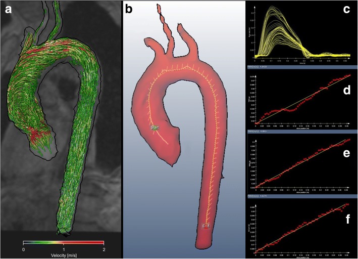
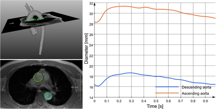
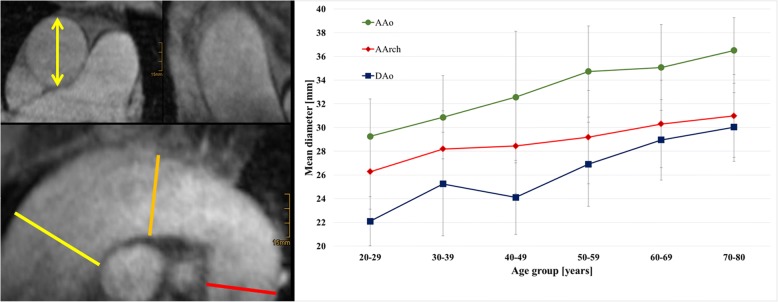
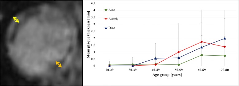
One hundred twenty six inhabitants of Freiburg, Germany, between 20 and 80 years prospectively underwent 3 Tesla cardiovascular magnetic resonance (CMR) of the thoracic aorta. 4D flow CMR (spatial/temporal resolution 2mm/20ms) was executed to calculate aortic pulse wave velocity (PWV) in m/s using dedicated software. In addition, we calculated distensibility coefficients (DC) using 2D CINE CMR imaging of the ascending (AAo) and descending aorta (DAo). Segmental aortic diameter and thickness of aortic plaques were determined by 3D T1 weighted CMR (spatial resolution 1mm).

RESULTS

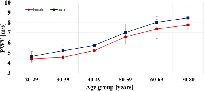
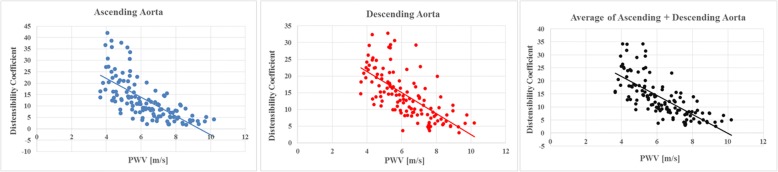
PWV increased from 4.93 ± 0.54 m/s in 20-30 year-old to 8.06 ± 1.03 m/s in 70-80 year-old subjects. PWV was significantly lower in women compared to men (p < 0.0001). Increased blood pressure (systolic r = 0.36, p < 0.0001; diastolic r = 0.33, p = 0.0001; mean arterial pressure r = 0.37, p < 0.0001) correlated with PWV after adjustment for age and gender. Finally, PWV increased with increasing diameter of the aorta (ascending aorta r = 0.20, p = 0.026; aortic arch r = 0.24, p = 0.009; descending aorta r = 0.26, p = 0.004). Correlation of PWV and DC of the AAo and DAo or the mean of both was high (r = 0.69, r = 0.68, r = 0.73; p < 0.001).

CONCLUSIONS

4D flow CMR was successfully applied to calculate aortic PWV and thus aortic stiffness. Findings showed a high correlation with distensibility coefficients representing local compliance of the aorta. Our novel method and reference data for PWV may provide a reliable biomarker for the identification of patients with underlying cardiovascular disease and optimal guidance of future treatment in studies or clinical routine.

摘要

背景

主动脉僵硬度增加是心血管疾病的独立预测因子。最佳测量方法对动脉粥样硬化的检测和高危患者的管理非常有益。因此,我们的目的是使用一种新的成像方法选择性地测量主动脉僵硬度,并从基于人群的研究中提供参考值。

方法

126 名德国弗赖堡居民前瞻性地接受了 3T 心脏磁共振(CMR)的胸主动脉检查。使用专用软件通过 4D 流量 CMR(空间/时间分辨率为 2mm/20ms)计算主动脉脉搏波速度(PWV),以 m/s 表示。此外,我们还使用升主动脉(AAo)和降主动脉(DAo)的 2D CINE CMR 成像计算了顺应性系数(DC)。通过 3D T1 加权 CMR(空间分辨率为 1mm)确定节段性主动脉直径和主动脉斑块厚度。

结果

PWV 从 20-30 岁的 4.93±0.54m/s 增加到 70-80 岁的 8.06±1.03m/s。女性的 PWV 明显低于男性(p<0.0001)。血压升高(收缩压 r=0.36,p<0.0001;舒张压 r=0.33,p=0.0001;平均动脉压 r=0.37,p<0.0001)与年龄和性别调整后的 PWV 相关。最后,PWV 随着主动脉直径的增加而增加(升主动脉 r=0.20,p=0.026;主动脉弓 r=0.24,p=0.009;降主动脉 r=0.26,p=0.004)。PWV 与 AAo 和 DAo 的 DC 或两者的平均值之间存在高度相关性(r=0.69,r=0.68,r=0.73;p<0.001)。

结论

4D 流量 CMR 成功地用于计算主动脉 PWV,从而测量主动脉僵硬度。结果与代表主动脉局部顺应性的顺应性系数高度相关。我们的新方法和 PWV 的参考数据可能为识别有潜在心血管疾病的患者提供可靠的生物标志物,并为研究或临床常规中的未来治疗提供最佳指导。

https://cdn.ncbi.nlm.nih.gov/pmc/blobs/3fab/6011486/fc85adac036a/12968_2018_461_Fig7_HTML.jpg
https://cdn.ncbi.nlm.nih.gov/pmc/blobs/3fab/6011486/249da83d2222/12968_2018_461_Fig1_HTML.jpg
https://cdn.ncbi.nlm.nih.gov/pmc/blobs/3fab/6011486/6cc20f6a274d/12968_2018_461_Fig2_HTML.jpg
https://cdn.ncbi.nlm.nih.gov/pmc/blobs/3fab/6011486/ac9b0f9ce457/12968_2018_461_Fig3_HTML.jpg
https://cdn.ncbi.nlm.nih.gov/pmc/blobs/3fab/6011486/538a497d4966/12968_2018_461_Fig4_HTML.jpg
https://cdn.ncbi.nlm.nih.gov/pmc/blobs/3fab/6011486/1e9002a4b063/12968_2018_461_Fig5_HTML.jpg
https://cdn.ncbi.nlm.nih.gov/pmc/blobs/3fab/6011486/86a080513ce1/12968_2018_461_Fig6_HTML.jpg
https://cdn.ncbi.nlm.nih.gov/pmc/blobs/3fab/6011486/fc85adac036a/12968_2018_461_Fig7_HTML.jpg

相似文献

1
Determination of aortic stiffness using 4D flow cardiovascular magnetic resonance - a population-based study.使用 4D 流心血管磁共振测定主动脉僵硬度 - 一项基于人群的研究。
J Cardiovasc Magn Reson. 2018 Jun 21;20(1):43. doi: 10.1186/s12968-018-0461-z.
2
Comparison of different methods for the estimation of aortic pulse wave velocity from 4D flow cardiovascular magnetic resonance.从 4D 流心血管磁共振评估主动脉脉搏波速度的不同方法比较。
J Cardiovasc Magn Reson. 2019 Dec 12;21(1):75. doi: 10.1186/s12968-019-0584-x.
3
Retrograde aortic blood flow as a mechanism of stroke: MR evaluation of the prevalence in a population-based study.逆行性主动脉血流是卒中的一种机制:基于人群的研究中其患病率的磁共振评估。
Eur Radiol. 2019 Oct;29(10):5172-5179. doi: 10.1007/s00330-019-06104-z. Epub 2019 Mar 15.
4
Simultaneous measurements of 3D wall shear stress and pulse wave velocity in the murine aortic arch.在小鼠主动脉弓中同时测量三维壁切应力和脉搏波速度。
J Cardiovasc Magn Reson. 2021 Mar 18;23(1):34. doi: 10.1186/s12968-021-00725-4.
5
Bramwell-Hill modeling for local aortic pulse wave velocity estimation: a validation study with velocity-encoded cardiovascular magnetic resonance and invasive pressure assessment.布拉姆韦尔-希尔模型用于局部主动脉脉搏波速度估计:一项基于速度编码心血管磁共振和有创压力评估的验证研究。
J Cardiovasc Magn Reson. 2012 Jan 9;14(1):2. doi: 10.1186/1532-429X-14-2.
6
Normal values of aortic dimensions, distensibility, and pulse wave velocity in children and young adults: a cross-sectional study.儿童和青年成年人的主动脉尺寸、可扩张性和脉搏波速度的正常值:一项横断面研究。
J Cardiovasc Magn Reson. 2012 Nov 14;14(1):77. doi: 10.1186/1532-429X-14-77.
7
Investigation of Aortic Wall Thickness, Stiffness and Flow Reversal in Patients With Cryptogenic Stroke: A 4D Flow MRI Study.隐源性卒中患者主动脉壁厚度、硬度及血流逆转的研究:一项四维血流磁共振成像研究
J Magn Reson Imaging. 2021 Mar;53(3):942-952. doi: 10.1002/jmri.27345. Epub 2020 Aug 31.
8
Quantification of aortic stiffness in stroke patients using 4D flow MRI in comparison with transesophageal echocardiography.应用 4D 血流 MRI 与经食管超声心动图对比评估脑卒中患者的主动脉僵硬度。
Int J Cardiovasc Imaging. 2018 Oct;34(10):1629-1636. doi: 10.1007/s10554-018-1369-2. Epub 2018 May 24.
9
Aortic length measurements for pulse wave velocity calculation: manual 2D vs automated 3D centreline extraction.用于脉搏波速度计算的主动脉长度测量:手动二维与自动三维中心线提取
J Cardiovasc Magn Reson. 2017 Mar 8;19(1):32. doi: 10.1186/s12968-017-0341-y.
10
Pediatric heterozygous familial hypercholesterolemia patients have locally increased aortic pulse wave velocity and wall thickness at the aortic root.儿科杂合子家族性高胆固醇血症患者的主动脉脉搏波速度和主动脉根部壁厚度局部增加。
Int J Cardiovasc Imaging. 2019 Oct;35(10):1903-1911. doi: 10.1007/s10554-019-01626-5. Epub 2019 Jun 17.

引用本文的文献

1
Virtual Therapy Planning of Aortic Valve Replacement for Preventing Patient-Prosthesis Mismatch.用于预防人工瓣膜与患者不匹配的主动脉瓣置换术的虚拟治疗规划
Bioengineering (Basel). 2025 Mar 21;12(4):328. doi: 10.3390/bioengineering12040328.
2
Variations in stiffness and structure of the human aorta along its length.人体主动脉沿其长度方向在硬度和结构上的变化。
Sci Rep. 2025 Apr 1;15(1):11120. doi: 10.1038/s41598-025-95008-8.
3
Male-female differences in thoracic aortic diameters at presentation of acute type A aortic dissection.急性A型主动脉夹层发病时胸主动脉直径的男女差异。

本文引用的文献

1
Ten-year longitudinal change in aortic stiffness assessed by cardiac MRI in the second half of the human lifespan: the multi-ethnic study of atherosclerosis.在人类寿命后半段通过心脏磁共振成像评估主动脉僵硬度的十年纵向变化:动脉粥样硬化的多民族研究
Eur Heart J Cardiovasc Imaging. 2016 Sep;17(9):1044-53. doi: 10.1093/ehjci/jev332. Epub 2016 Jan 12.
2
The two faces of hypertension: role of aortic stiffness.高血压的两面性:主动脉僵硬度的作用
J Am Soc Hypertens. 2016 Feb;10(2):175-83. doi: 10.1016/j.jash.2015.11.012. Epub 2015 Nov 26.
3
4D flow cardiovascular magnetic resonance consensus statement.
Int J Cardiol Heart Vasc. 2023 Oct 29;49:101290. doi: 10.1016/j.ijcha.2023.101290. eCollection 2023 Dec.
4
Paravascular fluid dynamics reveal arterial stiffness assessed using dynamic diffusion-weighted imaging.血管周围流体动力学揭示了使用动态扩散加权成像评估的动脉僵硬度。
NMR Biomed. 2024 Feb;37(2):e5048. doi: 10.1002/nbm.5048. Epub 2023 Oct 5.
5
Interpolation time-optimized aortic pulse wave velocity estimation by 4D flow MRI.基于 4D 流 MRI 的插值时间优化的主动脉脉搏波速度估计。
Sci Rep. 2023 Sep 30;13(1):16484. doi: 10.1038/s41598-023-43799-z.
6
Association between estimated pulse wave velocity and all-cause mortality in patients with coronary artery disease: a cohort study from NHANES 2005-2008.冠状动脉疾病患者脉搏波速度与全因死亡率的相关性:来自 NHANES 2005-2008 的队列研究。
BMC Cardiovasc Disord. 2023 Aug 21;23(1):412. doi: 10.1186/s12872-023-03435-0.
7
Aortic Stiffness Measured from Either 2D/4D Flow and Cine MRI or Applanation Tonometry in Coronary Artery Disease: A Case-Control Study.通过二维/四维血流和电影磁共振成像或压平眼压测量法测量冠状动脉疾病患者的主动脉僵硬度:一项病例对照研究。
J Clin Med. 2023 May 24;12(11):3643. doi: 10.3390/jcm12113643.
8
Magnetic Resonance Imaging and Computed Tomography for the Noninvasive Assessment of Arterial Aging: A Review by the VascAgeNet COST Action.磁共振成像和计算机断层扫描在动脉老化无创评估中的应用:VascAgeNet COST 行动综述。
J Am Heart Assoc. 2023 May 16;12(10):e027414. doi: 10.1161/JAHA.122.027414. Epub 2023 May 15.
9
Radiomics-based aortic flow profile characterization with 4D phase-contrast MRI.基于影像组学的4D相位对比磁共振成像对主动脉血流剖面的特征分析
Front Cardiovasc Med. 2023 Apr 3;10:1102502. doi: 10.3389/fcvm.2023.1102502. eCollection 2023.
10
Characterization of aortic aging using 3D multi-parametric MRI-long-term follow-up in a population study.使用 3D 多参数 MRI 对主动脉老化进行特征描述——一项人群研究的长期随访结果。
Sci Rep. 2023 Apr 18;13(1):6285. doi: 10.1038/s41598-023-33219-7.
4D 流动心血管磁共振共识声明。
J Cardiovasc Magn Reson. 2015 Aug 10;17(1):72. doi: 10.1186/s12968-015-0174-5.
4
Recommendations for Improving and Standardizing Vascular Research on Arterial Stiffness: A Scientific Statement From the American Heart Association.改善和规范动脉僵硬度血管研究的建议:美国心脏协会科学声明
Hypertension. 2015 Sep;66(3):698-722. doi: 10.1161/HYP.0000000000000033. Epub 2015 Jul 9.
5
Gender specific patterns of age-related decline in aortic stiffness: a cardiovascular magnetic resonance study including normal ranges.主动脉僵硬度随年龄增长的性别特异性模式:一项包含正常范围的心血管磁共振研究。
J Cardiovasc Magn Reson. 2015 Feb 19;17(1):20. doi: 10.1186/s12968-015-0126-0.
6
Proximal aortic distensibility is an independent predictor of all-cause mortality and incident CV events: the MESA study.升主动脉扩张性是全因死亡率和心血管事件发生率的独立预测因子:MESA 研究。
J Am Coll Cardiol. 2014 Dec 23;64(24):2619-2629. doi: 10.1016/j.jacc.2014.09.060.
7
Pulse wave velocity with 4D flow MRI: systematic differences and age-related regional vascular stiffness.基于四维血流磁共振成像的脉搏波速度:系统差异及与年龄相关的局部血管僵硬度
Magn Reson Imaging. 2014 Dec;32(10):1266-71. doi: 10.1016/j.mri.2014.08.021. Epub 2014 Aug 27.
8
Accelerated analysis of three-dimensional blood flow of the thoracic aorta in stroke patients.中风患者胸主动脉三维血流的加速分析
Int J Cardiovasc Imaging. 2014 Dec;30(8):1571-7. doi: 10.1007/s10554-014-0511-z. Epub 2014 Aug 15.
9
Aortic stiffness: pathophysiology, clinical implications, and approach to treatment.主动脉僵硬度:病理生理学、临床意义及治疗方法
Integr Blood Press Control. 2014 May 23;7:29-34. doi: 10.2147/IBPC.S59535. eCollection 2014.
10
Review of MRI-based measurements of pulse wave velocity: a biomarker of arterial stiffness.基于磁共振成像的脉搏波速度测量综述:动脉僵硬度的生物标志物
Cardiovasc Diagn Ther. 2014 Apr;4(2):193-206. doi: 10.3978/j.issn.2223-3652.2014.03.04.