• 文献检索
  • 文档翻译
  • 深度研究
  • 学术资讯
  • Suppr Zotero 插件Zotero 插件
  • 邀请有礼
  • 套餐&价格
  • 历史记录
应用&插件
Suppr Zotero 插件Zotero 插件浏览器插件Mac 客户端Windows 客户端微信小程序
定价
高级版会员购买积分包购买API积分包
服务
文献检索文档翻译深度研究API 文档MCP 服务
关于我们
关于 Suppr公司介绍联系我们用户协议隐私条款
关注我们

Suppr 超能文献

核心技术专利:CN118964589B侵权必究
粤ICP备2023148730 号-1Suppr @ 2026

文献检索

告别复杂PubMed语法,用中文像聊天一样搜索,搜遍4000万医学文献。AI智能推荐,让科研检索更轻松。

立即免费搜索

文件翻译

保留排版,准确专业,支持PDF/Word/PPT等文件格式,支持 12+语言互译。

免费翻译文档

深度研究

AI帮你快速写综述,25分钟生成高质量综述,智能提取关键信息,辅助科研写作。

立即免费体验

最近在非侵入性磁共振成像评估肝细胞癌方面的进展。

Recent advances in non-invasive magnetic resonance imaging assessment of hepatocellular carcinoma.

机构信息

School of Medicine, University of Milano-Bicocca, Milan 20126, Italy.

Division of Interventional Radiology, Department of Radiology, Madonna delle Grazie Hospital, Matera 75100, Italy.

出版信息

World J Gastroenterol. 2018 Jun 21;24(23):2413-2426. doi: 10.3748/wjg.v24.i23.2413.

DOI:10.3748/wjg.v24.i23.2413
PMID:29930464
原文链接:https://pmc.ncbi.nlm.nih.gov/articles/PMC6010944/
Abstract

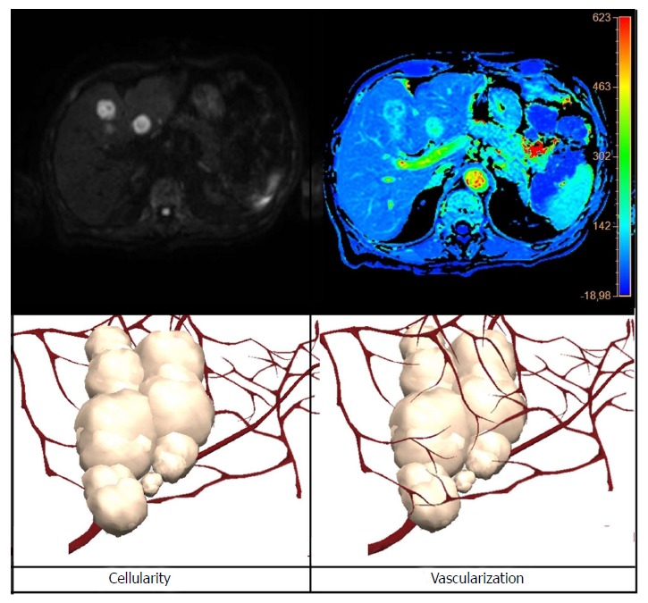
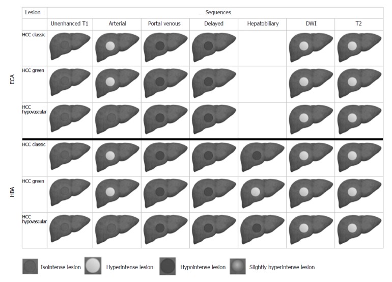
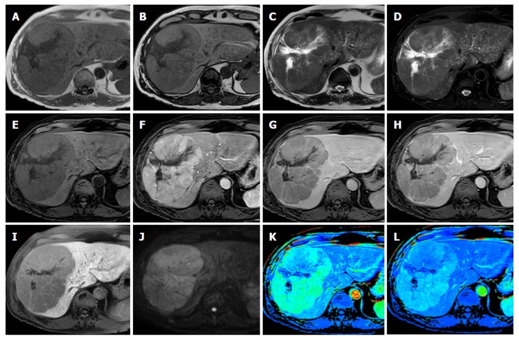
Magnetic resonance (MR) imaging of the liver is an important tool for the detection and characterization of focal liver lesions and for assessment of diffuse liver disease, having several intrinsic characteristics, represented by high soft tissue contrast, avoidance of ionizing radiation or iodinated contrast media, and more recently, by application of several functional imaging techniques (., diffusion-weighted sequences, hepatobiliary contrast agents, perfusion imaging, magnetic resonance (MR)-elastography, and radiomics analysis). MR functional imaging techniques are extensively used both in routine practice and in the field of clinical and pre-clinical research because, through a qualitative rather than quantitative approach, they can offer valuable information about tumor tissue and tissue architecture, cellular biomarkers related to the hepatocellular functions, or tissue vascularization profiles related to tumor and tissue biology. This kind of approach offers physiological parameters, capable of evaluating physiological and pathological modifications of tissues, by the analysis of quantitative data that could be used in tumor detection, characterization, treatment selection, and follow-up, in addition to those obtained from standard morphological imaging. In this review we provide an overview of recent advanced techniques in MR for the diagnosis and staging of hepatocellular carcinoma, and their role in the assessment of response treatment evaluation.

摘要

肝脏磁共振(MR)成像是检测和定性局灶性肝脏病变以及评估弥漫性肝脏疾病的重要工具,具有多种固有特征,包括高软组织对比度、避免电离辐射或碘造影剂,以及最近应用的多种功能成像技术(例如,扩散加权序列、肝胆对比剂、灌注成像、磁共振(MR)弹性成像和放射组学分析)。MR 功能成像技术在常规实践以及临床和临床前研究领域都得到了广泛应用,因为它们通过定性而非定量的方法可以提供有关肿瘤组织和组织结构、与肝细胞功能相关的细胞生物标志物或与肿瘤和组织生物学相关的组织血管化特征的有价值信息。这种方法提供了生理参数,可以通过分析可用于肿瘤检测、定性、治疗选择和随访的定量数据来评估组织的生理和病理变化,除了从标准形态成像获得的参数之外。在这篇综述中,我们概述了肝脏 MR 诊断和分期肝细胞癌的最新高级技术及其在评估治疗反应中的作用。

https://cdn.ncbi.nlm.nih.gov/pmc/blobs/a5c4/6010944/c412548c63cd/WJG-24-2413-g004.jpg
https://cdn.ncbi.nlm.nih.gov/pmc/blobs/a5c4/6010944/4873eeb3dd90/WJG-24-2413-g001.jpg
https://cdn.ncbi.nlm.nih.gov/pmc/blobs/a5c4/6010944/dee1d46ba570/WJG-24-2413-g002.jpg
https://cdn.ncbi.nlm.nih.gov/pmc/blobs/a5c4/6010944/f93c826f8ffd/WJG-24-2413-g003.jpg
https://cdn.ncbi.nlm.nih.gov/pmc/blobs/a5c4/6010944/c412548c63cd/WJG-24-2413-g004.jpg

相似文献

1
Recent advances in non-invasive magnetic resonance imaging assessment of hepatocellular carcinoma.最近在非侵入性磁共振成像评估肝细胞癌方面的进展。
World J Gastroenterol. 2018 Jun 21;24(23):2413-2426. doi: 10.3748/wjg.v24.i23.2413.
2
Noninvasive imaging of hepatocellular carcinoma: From diagnosis to prognosis.肝细胞癌的无创性影像学诊断与预后评估。
World J Gastroenterol. 2018 Jun 14;24(22):2348-2362. doi: 10.3748/wjg.v24.i22.2348.
3
Bifocal hepatocellular carcinoma: Magnetic resonance imaging features after transarterial embolization.双灶性肝细胞癌:经动脉栓塞后的磁共振成像特征
Curr Probl Cancer. 2018 May-Jun;42(3):319-321. doi: 10.1016/j.currproblcancer.2018.03.002. Epub 2018 Mar 28.
4
Advanced imaging techniques in the therapeutic response of transarterial chemoembolization for hepatocellular carcinoma.先进成像技术在肝细胞癌经动脉化疗栓塞治疗反应中的应用
World J Gastroenterol. 2016 May 28;22(20):4835-47. doi: 10.3748/wjg.v22.i20.4835.
5
Tumour-related neoangiogenesis: functional dynamic perfusion computed tomography for diagnosis and treatment efficacy assessment in hepatocellular carcinoma.肿瘤相关新生血管生成:功能动态灌注计算机断层扫描在肝细胞癌诊断及治疗疗效评估中的应用
Dig Liver Dis. 2014 Oct;46(10):916-22. doi: 10.1016/j.dld.2014.06.002. Epub 2014 Jul 9.
6
MRI in the evaluation of hepatocellular nodules: role of pulse sequences and contrast agents.磁共振成像在肝细胞结节评估中的应用:脉冲序列及对比剂的作用
Intervirology. 2004;47(3-5):252-70. doi: 10.1159/000078477.
7
New imaging techniques for liver diseases.肝脏疾病的新型影像学技术。
J Hepatol. 2015 Mar;62(3):690-700. doi: 10.1016/j.jhep.2014.10.014. Epub 2014 Oct 17.
8
The role of functional MR imaging in the assessment of tumor response after chemoembolization in patients with hepatocellular carcinoma.功能磁共振成像在评估肝细胞癌患者化疗栓塞术后肿瘤反应中的作用。
J Vasc Interv Radiol. 2006 Mar;17(3):505-12. doi: 10.1097/01.RVI.0000200052.02183.92.
9
Detection and characterization of focal liver lesions: a Japanese phase III, multicenter comparison between gadoxetic acid disodium-enhanced magnetic resonance imaging and contrast-enhanced computed tomography predominantly in patients with hepatocellular carcinoma and chronic liver disease.检测和描述肝脏局灶性病变:一项日本 III 期、多中心的钆塞酸二钠增强磁共振成像与增强计算机断层扫描对比研究,主要针对肝细胞癌和慢性肝病患者。
Invest Radiol. 2010 Mar;45(3):133-41. doi: 10.1097/RLI.0b013e3181caea5b.
10
Image fusion with volume navigation of contrast enhanced ultrasound (CEUS) with computed tomography (CT) or magnetic resonance imaging (MRI) for post-interventional follow-up after transcatheter arterial chemoembolization (TACE) of hepatocellular carcinomas (HCC): Preliminary results.超声造影(CEUS)容积导航图像融合与计算机断层扫描(CT)或磁共振成像(MRI)在肝癌(HCC)经导管动脉化疗栓塞(TACE)后的介入后随访中的应用:初步结果。
Clin Hemorheol Microcirc. 2010;46(2-3):101-15. doi: 10.3233/CH-2010-1337.

引用本文的文献

1
Radiomics analysis of R2* maps to predict early recurrence of single hepatocellular carcinoma after hepatectomy.基于R2*图的影像组学分析预测肝细胞癌肝切除术后早期复发
Front Oncol. 2024 Feb 23;14:1277698. doi: 10.3389/fonc.2024.1277698. eCollection 2024.
2
Role of gadoxetic acid-enhanced liver magnetic resonance imaging in the evaluation of hepatocellular carcinoma after locoregional treatment.钆塞酸增强肝脏磁共振成像在局部治疗后肝细胞癌评估中的作用。
World J Gastroenterol. 2022 Jul 14;28(26):3116-3131. doi: 10.3748/wjg.v28.i26.3116.
3
Current Imaging Diagnosis of Hepatocellular Carcinoma.

本文引用的文献

1
Quantitative free-breathing dynamic contrast-enhanced MRI in hepatocellular carcinoma using gadoxetic acid: correlations with Ki67 proliferation status, histological grades, and microvascular density.使用钆塞酸行定量自由呼吸动态对比增强 MRI 对肝细胞癌的研究:与 Ki67 增殖状态、组织学分级和微血管密度的相关性。
Abdom Radiol (NY). 2018 Jun;43(6):1393-1403. doi: 10.1007/s00261-017-1320-3.
2
Liver MRI: From basic protocol to advanced techniques.肝脏磁共振成像:从基础协议到先进技术。
Eur J Radiol. 2017 Aug;93:30-39. doi: 10.1016/j.ejrad.2017.05.028. Epub 2017 May 25.
3
How to Differentiate Borderline Hepatic Nodules in Hepatocarcinogenesis: Emphasis on Imaging Diagnosis.
肝细胞癌的当前影像诊断
Cancers (Basel). 2022 Aug 18;14(16):3997. doi: 10.3390/cancers14163997.
4
Benign focal liver lesions: The role of magnetic resonance imaging.肝脏良性局灶性病变:磁共振成像的作用
World J Hepatol. 2022 May 27;14(5):923-943. doi: 10.4254/wjh.v14.i5.923.
5
Surveillance of Fontan-associated liver disease: current standards and a proposal from the European Society of Paediatric Radiology Abdominal Task Force.法洛四联症相关肝病的监测:现行标准及欧洲儿科放射学会腹部工作组的建议。
Pediatr Radiol. 2021 Dec;51(13):2598-2606. doi: 10.1007/s00247-021-05173-x. Epub 2021 Oct 15.
6
Radiomics Analysis Based on Contrast-Enhanced MRI for Prediction of Therapeutic Response to Transarterial Chemoembolization in Hepatocellular Carcinoma.基于对比增强磁共振成像的放射组学分析预测肝细胞癌经动脉化疗栓塞术的治疗反应
Front Oncol. 2021 Mar 31;11:582788. doi: 10.3389/fonc.2021.582788. eCollection 2021.
7
Hepatic tumors: pitfall in diagnostic imaging.肝脏肿瘤:诊断影像学中的陷阱。
Acta Biomed. 2020 Jul 13;91(8-S):9-17. doi: 10.23750/abm.v91i8-S.9969.
8
Dynamic Hepatocellular Carcinoma Model Within a Liver Phantom for Multimodality Imaging.用于多模态成像的肝脏模型内动态肝细胞癌模型
Eur J Radiol Open. 2020 Sep 3;7:100257. doi: 10.1016/j.ejro.2020.100257. eCollection 2020.
9
Gadoxetic acid magnetic-enhanced resonance imaging in the diagnosis of cholangiocarcinoma.钆塞酸磁共振成像在胆管癌诊断中的应用。
World J Gastroenterol. 2020 Aug 7;26(29):4261-4271. doi: 10.3748/wjg.v26.i29.4261.
10
Role of dynamic perfusion magnetic resonance imaging in patients with local advanced rectal cancer.动态灌注磁共振成像在局部进展期直肠癌患者中的作用。
World J Gastroenterol. 2020 May 28;26(20):2657-2668. doi: 10.3748/wjg.v26.i20.2657.
肝癌发生过程中如何鉴别边缘性肝结节:重点在于影像诊断
Liver Cancer. 2017 Jun;6(3):189-203. doi: 10.1159/000455949. Epub 2017 Mar 9.
4
Diagnostic Performance of Gadoxetic Acid-enhanced Liver MR Imaging versus Multidetector CT in the Detection of Dysplastic Nodules and Early Hepatocellular Carcinoma.钆塞酸增强肝脏磁共振成像与多排 CT 在检测肝内不典型增生结节和早期肝细胞癌中的诊断性能比较。
Radiology. 2017 Oct;285(1):134-146. doi: 10.1148/radiol.2017162080. Epub 2017 Jun 13.
5
Image quality and diagnostic performance of free-breathing diffusion-weighted imaging for hepatocellular carcinoma.自由呼吸扩散加权成像对肝细胞癌的图像质量及诊断性能
World J Hepatol. 2017 May 18;9(14):657-666. doi: 10.4254/wjh.v9.i14.657.
6
Non-Hypervascular Hypointense Nodules at Gadoxetic Acid MRI: Hepatocellular Carcinoma Risk Assessment with Emphasis on the Role of Diffusion-Weighted Imaging.钆塞酸MRI上的非高血供低强化结节:以扩散加权成像的作用为重点的肝细胞癌风险评估
J Gastrointest Cancer. 2018 Sep;49(3):302-310. doi: 10.1007/s12029-017-9952-7.
7
Value of tumor stiffness measured with MR elastography for assessment of response of hepatocellular carcinoma to locoregional therapy.MR 弹性成像测量肿瘤硬度在评估肝癌局部治疗反应中的价值。
Abdom Radiol (NY). 2017 Jun;42(6):1685-1694. doi: 10.1007/s00261-017-1066-y.
8
Diffusion-weighted imaging (DWI) of hepatocellular carcinomas: a retrospective analysis of the correlation between qualitative and quantitative DWI and tumour grade.肝细胞癌的扩散加权成像(DWI):DWI定性和定量与肿瘤分级相关性的回顾性分析
Clin Radiol. 2017 Jun;72(6):465-472. doi: 10.1016/j.crad.2016.12.017. Epub 2017 Jan 19.
9
Early perfusion changes within 1 week of systemic treatment measured by dynamic contrast-enhanced MRI may predict survival in patients with advanced hepatocellular carcinoma.通过动态对比增强磁共振成像测量的全身治疗1周内的早期灌注变化可能预测晚期肝细胞癌患者的生存情况。
Eur Radiol. 2017 Jul;27(7):3069-3079. doi: 10.1007/s00330-016-4670-2. Epub 2016 Dec 12.
10
MR elastography of hepatocellular carcinoma: Correlation of tumor stiffness with histopathology features-Preliminary findings.肝细胞癌的磁共振弹性成像:肿瘤硬度与组织病理学特征的相关性——初步研究结果
Magn Reson Imaging. 2017 Apr;37:41-45. doi: 10.1016/j.mri.2016.11.005. Epub 2016 Nov 11.