Ahmad Muntaser S, Suardi Nursakinah, Shukri Ahmad, Nik Ab Razak Nik Noor Ashikin, Oglat Ammar A, Makhamrah Osama, Mohammad Hjouj
School of Physics, Universiti Sains Malaysia, 11800, Penang, Malaysia.
Department of Medical Imaging, Faculty of Applied Medical Sciences, Hashemite University, Zarqa, Jordan.
Eur J Radiol Open. 2020 Sep 3;7:100257. doi: 10.1016/j.ejro.2020.100257. eCollection 2020.
Hepatocellular carcinoma (HCC) is one of the most common cancer in the world, and the effectiveness of its treatment lies in its detection in its early stages. The aim of this study is to mimic HCC dynamically through a liver phantom and apply it in multimodality medical imaging techniques including magnetic resonance imaging (MRI), computed tomography (CT), and ultrasound.
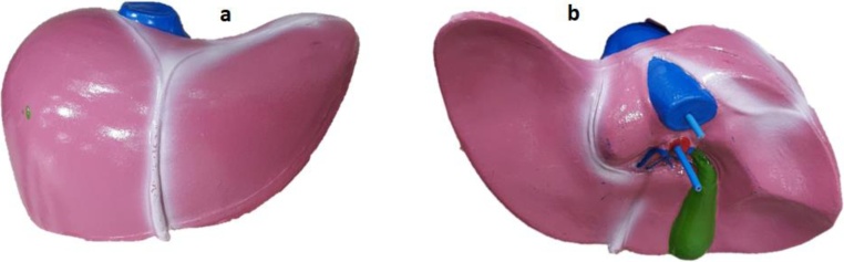
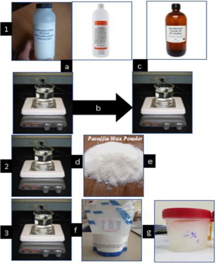
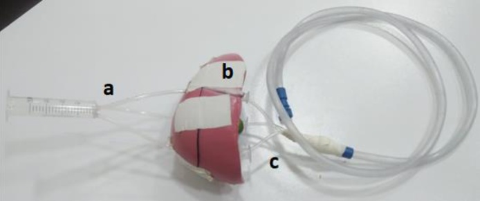
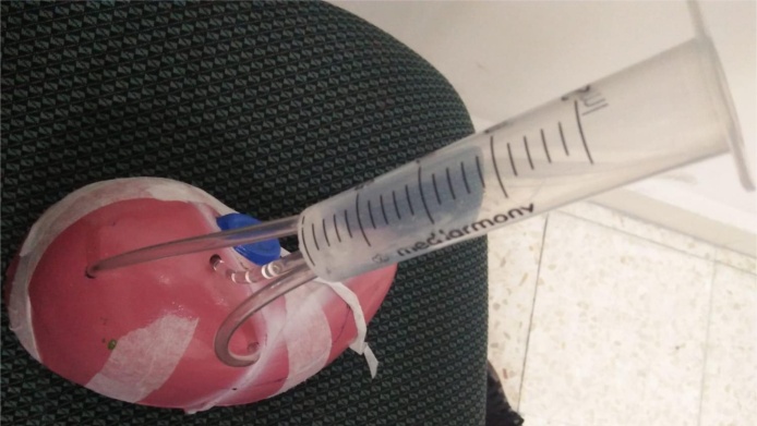
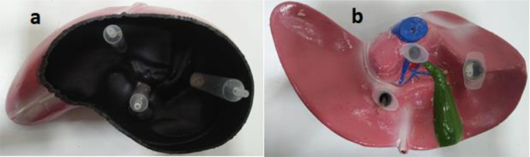
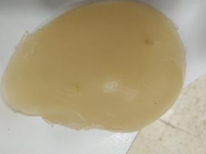
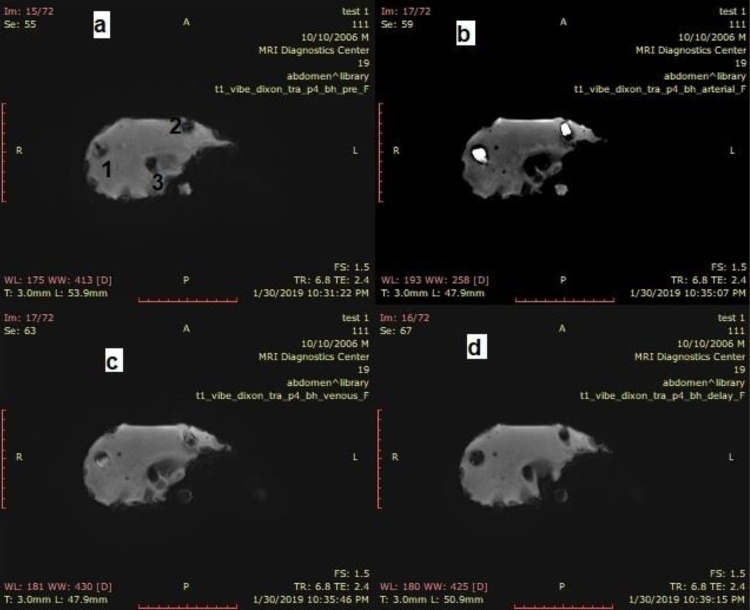
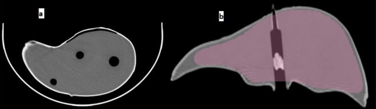
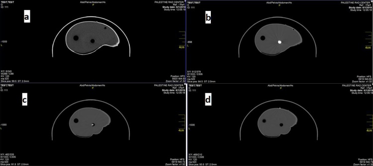
The phantom is fabricated with two main parts, liver parenchyma and HCC inserts. The liver parenchyma was fabricated by adding 2.5 wt% of agarose powder combined with 2.6 wt% of wax powder while the basic material for the HCC samples was made from polyurethane solution combined with 5 wt% glycerol. Three HCC samples were inserted into the parenchyma by using three cylinders implanted inside the liver parenchyma. An automatic injector is attached to the input side of the cylinders and a suction device connected to the output side of the cylinders. After the phantom was prepared, the contrast materials were injected into the phantom and imaged using MRI, CT, and ultrasound.
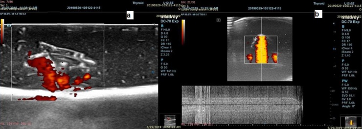
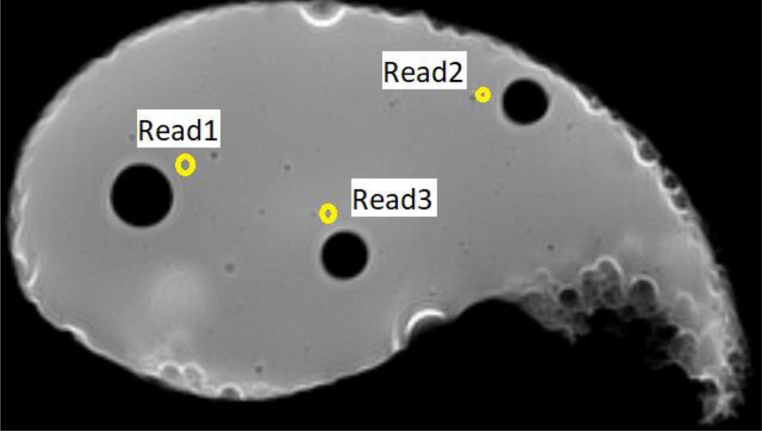
Both HCC samples and liver parenchyma were clearly distinguished using the three imaging modalities: MRI, CT, and ultrasound. Doppler ultrasound was also applied through the HCC samples and the flow pattern was observed through the samples.
A multimodal dynamic liver phantom, with HCC tumor models have been fabricated. This phantom helps to improve and develop different methods for detecting HCC in its early stages.
肝细胞癌(HCC)是世界上最常见的癌症之一,其治疗效果取决于早期检测。本研究的目的是通过肝脏模型动态模拟HCC,并将其应用于包括磁共振成像(MRI)、计算机断层扫描(CT)和超声在内的多模态医学成像技术。
该模型由两个主要部分制成,即肝实质和HCC植入物。肝实质是通过添加2.5 wt%的琼脂糖粉和2.6 wt%的蜡粉制成,而HCC样本的基础材料则由聚氨酯溶液和5 wt%的甘油制成。通过植入肝实质内的三个圆柱体将三个HCC样本插入实质中。一个自动注射器连接到圆柱体的输入端,一个抽吸装置连接到圆柱体的输出端。制备好模型后,将对比剂注入模型并使用MRI、CT和超声进行成像。
使用MRI、CT和超声这三种成像方式都能清晰区分HCC样本和肝实质。还通过HCC样本应用了多普勒超声,并观察了样本中的血流模式。
已制作出具有HCC肿瘤模型的多模态动态肝脏模型。该模型有助于改进和开发早期检测HCC的不同方法。