Department of Otolaryngology, Head and Neck Surgery, University Hospital of Basel, Basel, Switzerland.
Department of Biomedicine, University of Basel, Basel, Switzerland.
PLoS One. 2018 Jun 22;13(6):e0198029. doi: 10.1371/journal.pone.0198029. eCollection 2018.
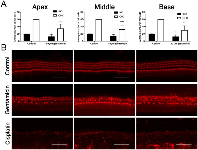
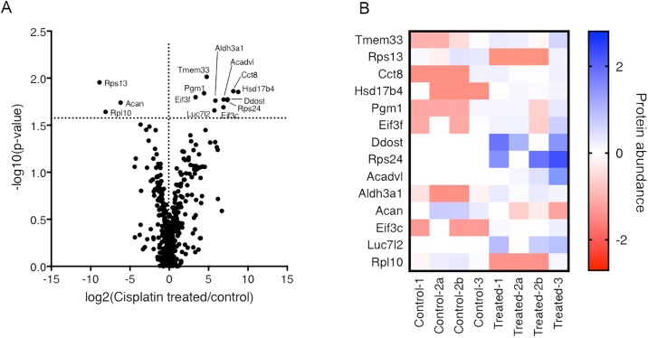
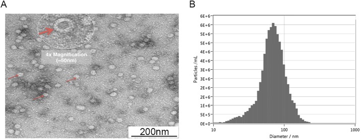
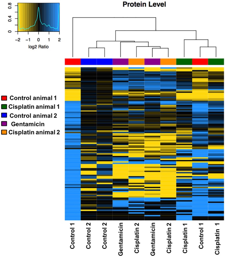
Exosomes are nanovesicles involved in intercellular communications. They are released by a variety of cell types; however, their presence in the inner ear has not been described in the literature. The aims of this study were to determine if exosomes are present in the inner ear and, if present, characterize the changes in their protein content in response to ototoxic stress. In this laboratory investigation, inner ear explants of 5-day-old Wistar rats were cultured and treated with either cisplatin or gentamicin. Hair cell damage was assessed by confocal microscopy. Exosomes were isolated using ExoQuick, serial centrifugation, and mini-column methods. Confirmation and characterization of exosomes was carried out using transmission electron microscopy (TEM), ZetaView, BCA protein analysis, and proteomics. Vesicles with a typical size distribution for exosomes were observed using TEM and ZetaView. Proteomic analysis detected typical exosome markers and markers for the organ of Corti. There was a statistically significant reduction in the exosome protein level and number of particles per cubic centimeter when the samples were exposed to ototoxic stress. Proteomic analysis also detected clear differences in protein expression when ototoxic medications were introduced. Significant changes in the proteomes of the exosomes were previously described in the context of hearing loss and ototoxic treatment. This is the first report describing exosomes derived from the inner ear. These findings may present an opportunity to conduct further studies with the hope of using exosomes as a biomarker to monitor inner ear function in the future.
外泌体是参与细胞间通讯的纳米囊泡。它们由多种细胞类型释放,但在内耳中尚未有文献描述其存在。本研究旨在确定外泌体是否存在于内耳中,如果存在,研究其蛋白质含量在外耳毒性应激下的变化特征。在这项实验室研究中,培养了 5 天大的 Wistar 大鼠内耳组织外植体,并分别用顺铂或庆大霉素处理。通过共聚焦显微镜评估毛细胞损伤。使用 ExoQuick、连续离心和迷你柱方法分离外泌体。使用透射电子显微镜(TEM)、ZetaView、BCA 蛋白分析和蛋白质组学对其进行确认和特征分析。TEM 和 ZetaView 观察到具有典型外泌体大小分布的囊泡。蛋白质组学分析检测到典型的外泌体标志物和 Corti 器的标志物。当样本暴露在外耳毒性应激下时,外泌体蛋白水平和每立方厘米颗粒数显著降低。蛋白质组学分析还检测到引入耳毒性药物时蛋白质表达的明显差异。在外耳毒性和耳毒性治疗的背景下,外泌体的蛋白质组发生显著变化已有报道。这是首次描述源自内耳的外泌体的报告。这些发现可能为进一步研究提供机会,以期将来使用外泌体作为监测内耳功能的生物标志物。