Department of Comparative Biology and Experimental Medicine, Faculty of Veterinary Medicine, University of Calgary, Calgary, Alberta T2N 4Z6, Canada; Department of Molecular Microbiology and Immunology, Division of Cellular and Molecular Biology, Graduate School of Biomedical Sciences, Nagasaki University, Nagasaki 852-8523, Japan.
Department of Comparative Biology and Experimental Medicine, Faculty of Veterinary Medicine, University of Calgary, Calgary, Alberta T2N 4Z6, Canada; Calgary Prion Research Unit, University of Calgary, Calgary, Alberta, Canada.
J Biol Chem. 2018 Aug 17;293(33):12730-12740. doi: 10.1074/jbc.RA117.001633. Epub 2018 Jun 22.
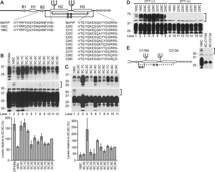
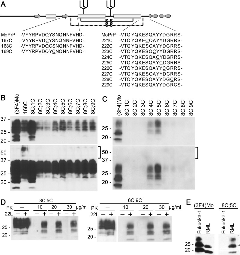
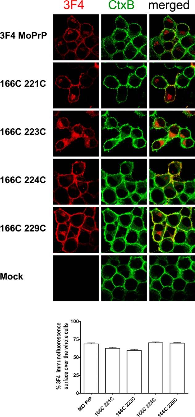
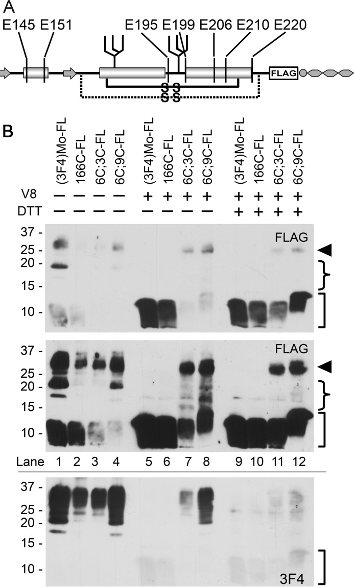
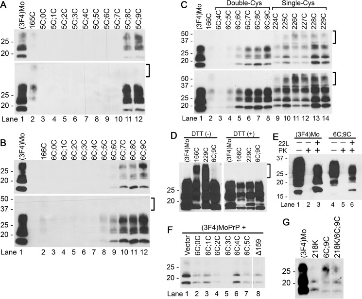
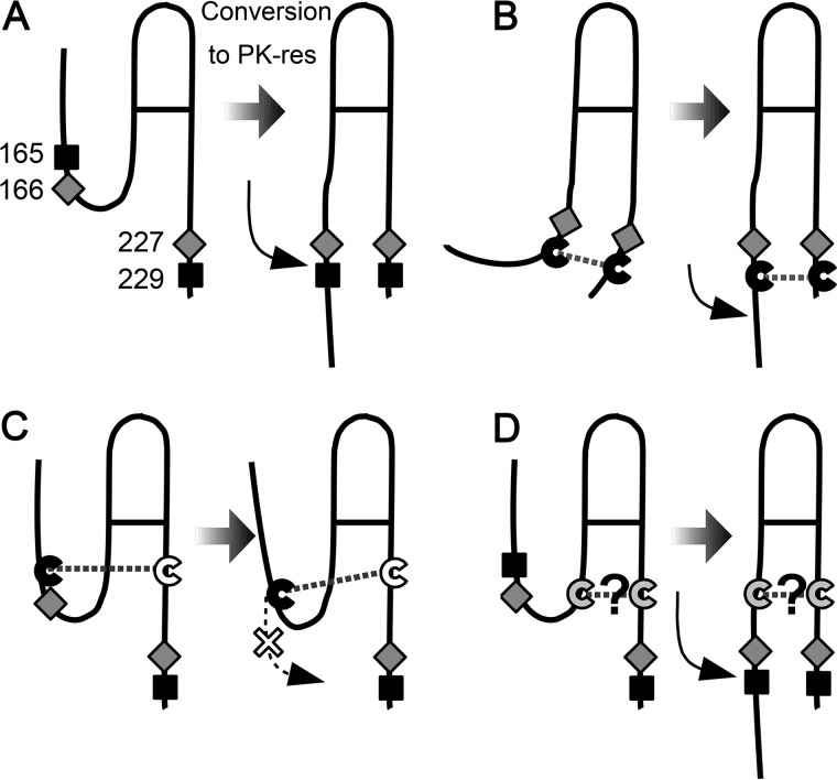
Prions are composed solely of the pathological isoform (PrP) of the normal cellular prion protein (PrP). Identification of different PrP structures is crucially important for understanding prion biology because the pathogenic properties of prions are hypothesized to be encoded in the structures of PrP However, these structures remain yet to be identified, because of the incompatibility of PrP with conventional high-resolution structural analysis methods. Previously, we reported that the region between the first and the second α-helix (H1∼H2) of PrP might cooperate with the more C-terminal side region for efficient interactions with PrP From this starting point, we created a series of PrP variants with two cysteine substitutions (C;C-PrP) forming a disulfide-crosslink between H1∼H2 and the distal region of the third helix (Ctrm). We then assessed the conversion capabilities of the C;C-PrP variants in N2a cells infected with mouse-adapted scrapie prions (22L-ScN2a). Specifically, Cys substitutions at residues 165, 166, or 168 in H1∼H2 were combined with cysteine scanning along Ctrm residues 220-229. We found that C;C-PrPs are expressed normally with glycosylation patterns and subcellular localization similar to WT PrP, albeit differing in expression levels. Interestingly, some C;C-PrPs converted to protease-resistant isoforms in the 22L-ScN2a cells, but not in Fukuoka1 prion-infected cells. Crosslink patterns of convertible C;C-PrPs indicated a positional change of H1∼H2 toward Ctrm in PrP-induced conformational conversion. Given the properties of the C;C-PrPs reported here, we propose that these PrP variants may be useful tools for investigating prion strain-specific structures and structure-phenotype relationships of PrP.
朊病毒仅由正常细胞朊病毒蛋白(PrP)的病理性异构体(PrP)组成。鉴定不同的 PrP 结构对于理解朊病毒生物学至关重要,因为假设朊病毒的致病性是由 PrP 的结构编码的。然而,由于 PrP 与传统的高分辨率结构分析方法不兼容,这些结构仍然有待确定。以前,我们报道过 PrP 的第一和第二α螺旋(H1∼H2)之间的区域可能与更 C 端的区域合作,以实现与 PrP 的有效相互作用。从这个起点出发,我们创建了一系列具有两个半胱氨酸取代(C;C-PrP)的 PrP 变体,在 H1∼H2 和第三螺旋(Ctrm)的远端区域之间形成二硫键交联。然后,我们评估了 C;C-PrP 变体在感染鼠源传染性海绵状脑病朊病毒(22L-ScN2a)的 N2a 细胞中的转化能力。具体而言,H1∼H2 中残基 165、166 或 168 的半胱氨酸取代与 Ctrm 残基 220-229 的半胱氨酸扫描相结合。我们发现 C;C-PrP 正常表达,糖基化模式和亚细胞定位与 WT PrP 相似,尽管表达水平不同。有趣的是,一些 C;C-PrP 在 22L-ScN2a 细胞中转化为蛋白酶抗性异构体,但在福冈 1 朊病毒感染的细胞中则没有。可转化的 C;C-PrP 的交联模式表明 H1∼H2 在 PrP 诱导的构象转换中向 Ctrm 的位置发生变化。鉴于这里报道的 C;C-PrP 的特性,我们提出这些 PrP 变体可能是研究朊病毒株特异性结构和 PrP 结构-表型关系的有用工具。