Taguchi Yuzuru, Schätzl Hermann M
Department of Comparative Biology & Experimental Medicine; Faculty of Veterinary Medicine; University of Calgary; Calgary, AB Canada.
Department of Comparative Biology & Experimental Medicine; Faculty of Veterinary Medicine; University of Calgary; Calgary, AB Canada; Departments of Molecular Biology and of Veterinary Sciences; University of Wyoming; Laramie, Wyoming, USA.
Prion. 2013 Nov-Dec;7(6):452-6. doi: 10.4161/pri.27500. Epub 2013 Dec 24.
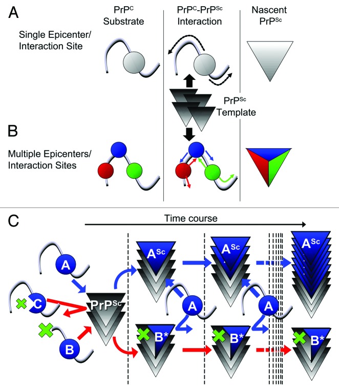
A direct physical interaction of the prion protein isoforms is a key element in prion conversion. Which sites interact first and which parts of PrP(c) are converted subsequently is presently not known in detail. We hypothesized that structural changes induced by PrP(Sc) interaction occur in more than one interface and subsequently propagate within the PrP(C) substrate, like epicenters of structural changes. To identify potential interfaces we created a series of systematically-designed mutant PrPs and tested them in prion-infected cells for dominant-negative inhibition (DNI) effects. This showed that mutant PrPs with deletions in the region between first and second α-helix are involved in PrP-PrP interaction and conversion of PrP(C) into PrP(Sc). Although some PrPs did not reach the plasma membrane, they had access to the locales of prion conversion and PrP(Sc) recycling using autophagy pathways. Using other series of mutant PrPs we already have identified additional sites which constitute potential interaction interfaces. Our approach has the potential to characterize PrP-PrP interaction sites in the context of prion-infected cells. Besides providing further insights into the molecular mechanisms of prion conversion, this data may help to further elucidate how prion strain diversity is maintained.
朊病毒蛋白异构体之间的直接物理相互作用是朊病毒转化的关键因素。目前尚不清楚哪些位点首先发生相互作用,以及随后PrP(c)的哪些部分会发生转化。我们推测,由PrP(Sc)相互作用诱导的结构变化发生在多个界面,并随后在PrP(C)底物内传播,就像结构变化的震中一样。为了识别潜在的界面,我们创建了一系列系统设计的突变型PrP,并在朊病毒感染的细胞中测试它们的显性负抑制(DNI)效应。结果表明,在第一个和第二个α-螺旋之间区域有缺失的突变型PrP参与了PrP-PrP相互作用以及PrP(C)向PrP(Sc)的转化。尽管一些PrP没有到达质膜,但它们可以通过自噬途径进入朊病毒转化和PrP(Sc)循环利用的场所。使用其他系列的突变型PrP,我们已经确定了构成潜在相互作用界面的其他位点。我们的方法有潜力在朊病毒感染的细胞背景下表征PrP-PrP相互作用位点。除了进一步深入了解朊病毒转化的分子机制外,这些数据可能有助于进一步阐明朊病毒株多样性是如何维持的。