International Research Center for Medical Sciences, Kumamoto University, Japan
International Research Center for Medical Sciences, Kumamoto University, Japan.
J Exp Med. 2018 Aug 6;215(8):2097-2113. doi: 10.1084/jem.20180421. Epub 2018 Jun 26.
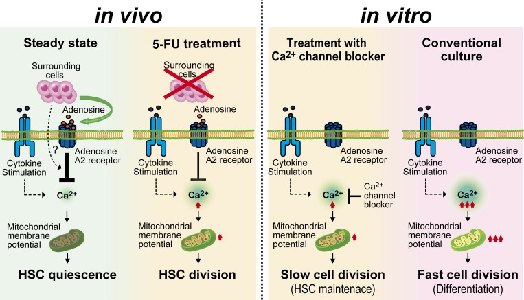
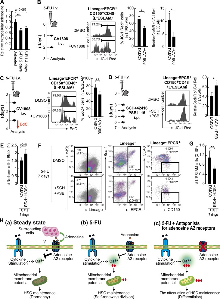
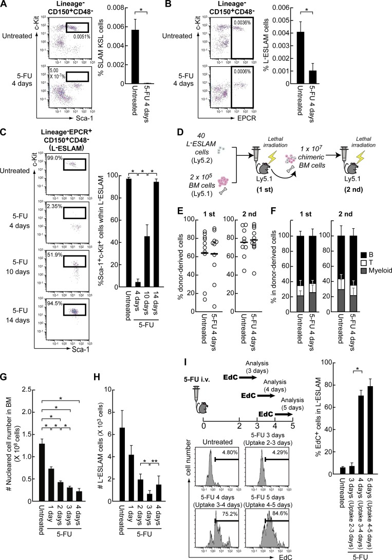
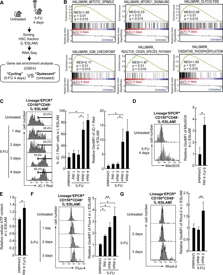
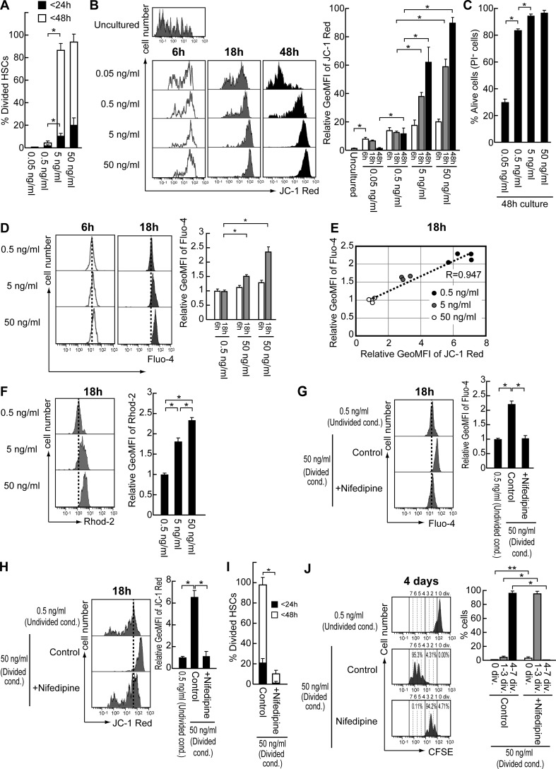
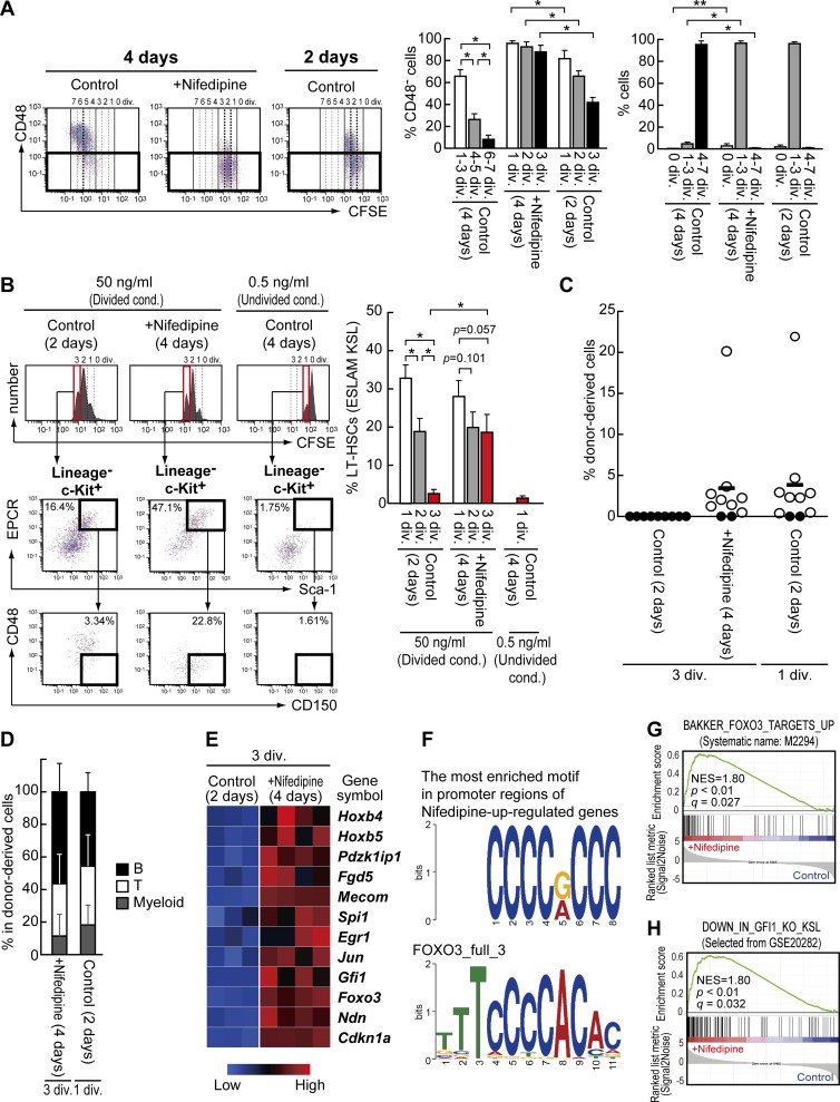
Most of the hematopoietic stem cells (HSCs) within the bone marrow (BM) show quiescent state with a low mitochondrial membrane potential (ΔΨ). In contrast, upon stress hematopoiesis, HSCs actively start to divide. However, the underlying mechanism for the initiation of HSC division still remains unclear. To elucidate the mechanism underlying the transition of cell cycle state in HSCs, we analyzed the change of mitochondria in HSCs after BM suppression induced by 5-fluoruracil (5-FU). We found that HSCs initiate cell division after exhibiting enhanced ΔΨ as a result of increased intracellular Ca level. Although further activation of Ca-mitochondria pathway led to loss of HSCs after cell division, the appropriate suppression of intracellular Ca level by exogenous adenosine or Nifedipine, a Ca channel blocker, prolonged cell division interval in HSCs, and simultaneously achieved both cell division and HSC maintenance. Collectively, our results indicate that the Ca-mitochondria pathway induces HSC division critically to determine HSC cell fate.
大多数骨髓(BM)中的造血干细胞(HSCs)处于静止状态,具有较低的线粒体膜电位(ΔΨ)。相比之下,在应激造血时,HSCs 会积极开始分裂。然而,HSC 分裂起始的潜在机制仍不清楚。为了阐明 HSCs 细胞周期状态转变的机制,我们分析了 5-氟尿嘧啶(5-FU)诱导 BM 抑制后 HSCs 中线粒体的变化。我们发现,HSCs 在细胞内 Ca 水平升高导致 ΔΨ 增强后开始细胞分裂。尽管 Ca-线粒体途径的进一步激活导致细胞分裂后 HSCs 的丢失,但通过外源性腺苷或 Ca 通道阻滞剂硝苯地平适当抑制细胞内 Ca 水平可延长 HSCs 的细胞分裂间隔,同时实现细胞分裂和 HSC 维持。总之,我们的结果表明 Ca-线粒体途径可诱导 HSC 分裂,从而决定 HSC 细胞命运。