Lopes César Vivian, Hartmann Antônio Atalíbio, Artifon Everson Luiz de Almeida
Department of Gastroenterology and Digestive Endoscopy.
Department of Pathology, Santa Casa Hospital, Porto Alegre, RS.
Arq Bras Cir Dig. 2018 Jun 21;31(1):e1350. doi: 10.1590/0102-672020180001e1350.
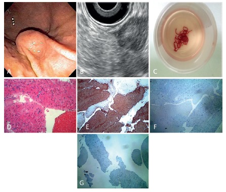
Tissue diagnosis is required for gastric subepithelial lesions for differential diagnosis of GISTs. However, there has not been consensus about the best needle for EUS-guided sampling of these lesions.
To evaluate the diagnostic yield of EUS-FNA for gastric subepithelial lesions of the proper muscle layer with large-bore 19 gauge needles.
A prospectively maintained database was retrospectively reviewed to identify consecutive patients who underwent EUS-FNA with 19 and 22 gauge needles for gastric subepithelial lesions of the fourth endosonographic layer in a tertiary care referral center. EUS-FNA was performed by the same endosonographer, using the fanning technique, without on-site cytopathologist. Specimens were analysed through cell blocks by the same pathologist. Procedure results were categorized into diagnostic, defined as enough material for histopathology and immunohistochemistry, or nondiagnostic.
Eighty-nine patients (mean age: 59 years, 77% women) underwent 92 EUS-FNA with 19 (75) or 22 (17) gauge needles. Mean lesion size was 22.6 mm. Overall diagnostic yield was 88%. The diagnostic yield of 19 gauge was higher than that of 22 gauge needle (92%x70.6%; p=0.0410), and similar for lesions >2 cm and <2 cm (93.7%x90.7%; p=0.9563). The best performance for 19 gauge needles was obtained performing <3 needle passes. Complication rate was 2.8%.
Diagnostic yield of EUS-FNA with 19 gauge needles is 92% for gastric subepithelial lesions of the proper muscle layer. It is safe and highly valuable for differentiation between GIST and leiomyoma, no matter the size of the lesion.
胃上皮下病变的组织诊断对于胃肠间质瘤(GIST)的鉴别诊断是必要的。然而,对于这些病变的超声内镜引导下采样的最佳针具尚无共识。
评估使用19G大口径针进行超声内镜引导下细针穿刺抽吸术(EUS-FNA)对胃固有肌层上皮下病变的诊断率。
回顾性分析一个前瞻性维护的数据库,以确定在一家三级医疗转诊中心接受19G和22G针超声内镜引导下细针穿刺抽吸术的连续患者,这些患者的胃上皮下病变位于超声内镜第四层。EUS-FNA由同一位超声内镜医生进行,采用扇形技术,且无现场细胞病理学家。标本由同一位病理学家通过细胞块进行分析。将操作结果分为诊断性(定义为有足够材料进行组织病理学和免疫组织化学检查)或非诊断性。
89例患者(平均年龄:59岁,77%为女性)接受了92次EUS-FNA,使用19G针75次,22G针17次。病变平均大小为22.6mm。总体诊断率为88%。19G针的诊断率高于22G针(92%对70.6%;p=0.0410),对于大于2cm和小于2cm的病变诊断率相似(93.7%对90.7%;p=0.9563)。19G针在穿刺针数<3针时表现最佳。并发症发生率为2.8%。
对于胃固有肌层上皮下病变,使用19G针进行EUS-FNA的诊断率为92%。无论病变大小,对于鉴别GIST和平滑肌瘤都是安全且非常有价值的。