• 文献检索
  • 文档翻译
  • 深度研究
  • 学术资讯
  • Suppr Zotero 插件Zotero 插件
  • 邀请有礼
  • 套餐&价格
  • 历史记录
应用&插件
Suppr Zotero 插件Zotero 插件浏览器插件Mac 客户端Windows 客户端微信小程序
定价
高级版会员购买积分包购买API积分包
服务
文献检索文档翻译深度研究API 文档MCP 服务
关于我们
关于 Suppr公司介绍联系我们用户协议隐私条款
关注我们

Suppr 超能文献

核心技术专利:CN118964589B侵权必究
粤ICP备2023148730 号-1Suppr @ 2026

文献检索

告别复杂PubMed语法,用中文像聊天一样搜索,搜遍4000万医学文献。AI智能推荐,让科研检索更轻松。

立即免费搜索

文件翻译

保留排版,准确专业,支持PDF/Word/PPT等文件格式,支持 12+语言互译。

免费翻译文档

深度研究

AI帮你快速写综述,25分钟生成高质量综述,智能提取关键信息,辅助科研写作。

立即免费体验

使用心血管磁共振基于钆对比前后 T1 映射计算心肌细胞外容积(ECV)时,标准化红细胞比容测量时间的重要性。

Importance of standardizing timing of hematocrit measurement when using cardiovascular magnetic resonance to calculate myocardial extracellular volume (ECV) based on pre- and post-contrast T1 mapping.

机构信息

Department of Clinical Physiology, Clinical Sciences, Lund University and Lund University Hospital, Getingevägen 3, 221 85, Lund, Sweden.

Laboratory of Computing, Medical Informatics and Biomedical - Imaging Technologies, School of Medicine, Aristotle University of Thessaloniki, Thessaloniki, Greece.

出版信息

J Cardiovasc Magn Reson. 2018 Jun 28;20(1):46. doi: 10.1186/s12968-018-0464-9.

DOI:10.1186/s12968-018-0464-9
PMID:29950178
原文链接:https://pmc.ncbi.nlm.nih.gov/articles/PMC6022290/
Abstract

BACKGROUND

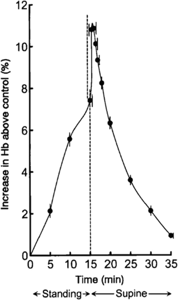
Cardiovascular magnetic resonance (CMR) can be used to calculate myocardial extracellular volume fraction (ECV) by relating the longitudinal relaxation rate in blood and myocardium before and after contrast-injection to hematocrit (Hct) in blood. Hematocrit is known to vary with body posture, which could affect the calculations of ECV. The aim of this study was to test the hypothesis that there is a significant increase in calculated ECV values if the Hct is sampled after the CMR examination in supine position compared to when the patient arrives at the MR department.

METHODS

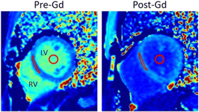
Forty-three consecutive patients including various pathologies as well as normal findings were included in the study. Venous blood samples were drawn upon arrival to the MR department and directly after the examination with the patient remaining in supine position. A Modified Look-Locker Inversion recovery (MOLLI) protocol was used to acquire mid-ventricular short-axis images before and after contrast injection from which motion-corrected T1 maps were derived and ECV was calculated.

RESULTS

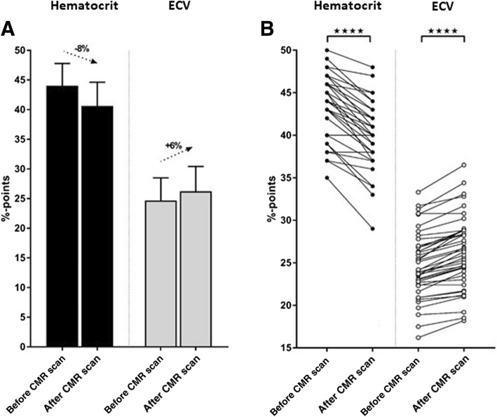
Hematocrit decreased from 44.0 ± 3.7% before to 40.6 ± 4.0% after the CMR examination (p < 0.001). This resulted in a change in calculated ECV from 24.7 ± 3.8% before to 26.2 ± 4.2% after the CMR examination (p < 0.001). All patients decreased in Hct after the CMR examination compared to before except for two patients whose Hct remained the same.

CONCLUSION

Variability in CMR-derived myocardial ECV can be reduced by standardizing the timing of Hct measurement relative to the CMR examination. Thus, a standardized acquisition of blood sample for Hct after the CMR examination, when the patient is still in supine position, would increase the precision of ECV measurements.

摘要

背景

心血管磁共振(CMR)可用于通过比较血中和心肌内对比注射前后的纵向弛豫率与血红细胞压积(Hct)来计算心肌细胞外容积分数(ECV)。已知红细胞压积会随体位变化而变化,这可能会影响 ECV 的计算。本研究旨在验证以下假设,即在患者处于仰卧位时进行 CMR 检查后采集 Hct 样本,与患者到达磁共振科时采集 Hct 样本相比,计算出的 ECV 值会显著增加。

方法

研究纳入了 43 例连续患者,包括各种病理和正常发现。患者到达磁共振科后和检查后立即采集静脉血样,此时患者保持仰卧位。采用改良 Look-Locker 反转恢复(MOLLI)序列在对比注射前后获取中室短轴图像,从这些图像中得出运动校正 T1 图,并计算 ECV。

结果

Hct 从 CMR 检查前的 44.0±3.7%降至检查后的 40.6±4.0%(p<0.001)。这导致计算出的 ECV 从 CMR 检查前的 24.7±3.8%变为检查后的 26.2±4.2%(p<0.001)。除了两名患者的 Hct 不变外,所有患者在 CMR 检查后都比检查前的 Hct 降低。

结论

通过使 Hct 测量的时间与 CMR 检查相对标准化,可以减少 CMR 衍生的心肌 ECV 的变异性。因此,当患者仍处于仰卧位时,标准化采集 CMR 检查后用于 Hct 的血液样本,将提高 ECV 测量的精度。

https://cdn.ncbi.nlm.nih.gov/pmc/blobs/873a/6022290/d1cf2ad8a6b7/12968_2018_464_Fig3_HTML.jpg
https://cdn.ncbi.nlm.nih.gov/pmc/blobs/873a/6022290/6f379365ecf3/12968_2018_464_Fig1_HTML.jpg
https://cdn.ncbi.nlm.nih.gov/pmc/blobs/873a/6022290/e05b323fa4e6/12968_2018_464_Fig2_HTML.jpg
https://cdn.ncbi.nlm.nih.gov/pmc/blobs/873a/6022290/d1cf2ad8a6b7/12968_2018_464_Fig3_HTML.jpg
https://cdn.ncbi.nlm.nih.gov/pmc/blobs/873a/6022290/6f379365ecf3/12968_2018_464_Fig1_HTML.jpg
https://cdn.ncbi.nlm.nih.gov/pmc/blobs/873a/6022290/e05b323fa4e6/12968_2018_464_Fig2_HTML.jpg
https://cdn.ncbi.nlm.nih.gov/pmc/blobs/873a/6022290/d1cf2ad8a6b7/12968_2018_464_Fig3_HTML.jpg

相似文献

1
Importance of standardizing timing of hematocrit measurement when using cardiovascular magnetic resonance to calculate myocardial extracellular volume (ECV) based on pre- and post-contrast T1 mapping.使用心血管磁共振基于钆对比前后 T1 映射计算心肌细胞外容积(ECV)时,标准化红细胞比容测量时间的重要性。
J Cardiovasc Magn Reson. 2018 Jun 28;20(1):46. doi: 10.1186/s12968-018-0464-9.
2
Automatic Measurement of the Myocardial Interstitium: Synthetic Extracellular Volume Quantification Without Hematocrit Sampling.心肌间质的自动测量:无需红细胞压积采样的合成细胞外体积定量。
JACC Cardiovasc Imaging. 2016 Jan;9(1):54-63. doi: 10.1016/j.jcmg.2015.11.008.
3
Is a timely assessment of the hematocrit necessary for cardiovascular magnetic resonance-derived extracellular volume measurements?是否需要及时评估血细胞比容以进行心血管磁共振衍生的细胞外容积测量?
J Cardiovasc Magn Reson. 2020 Nov 30;22(1):77. doi: 10.1186/s12968-020-00689-x.
4
Comprehensive validation of cardiovascular magnetic resonance techniques for the assessment of myocardial extracellular volume.全面验证心血管磁共振技术评估心肌细胞外容积。
Circ Cardiovasc Imaging. 2013 May 1;6(3):373-83. doi: 10.1161/CIRCIMAGING.112.000192. Epub 2013 Apr 3.
5
Extracellular volume fraction measurements derived from the longitudinal relaxation of blood-based synthetic hematocrit may lead to clinical errors in 3 T cardiovascular magnetic resonance.基于血液合成血细胞比容的纵向弛豫测量得出的细胞外容积分数可能会导致 3T 心血管磁共振中的临床错误。
J Cardiovasc Magn Reson. 2018 Aug 9;20(1):56. doi: 10.1186/s12968-018-0475-6.
6
T1 mapping of the myocardium: intra-individual assessment of post-contrast T1 time evolution and extracellular volume fraction at 3T for Gd-DTPA and Gd-BOPTA.心肌 T1 映射:3T 下钆-DTPA 和钆-BOPTA 对比后 T1 时间演变和细胞外容积分数的个体内评估。
J Cardiovasc Magn Reson. 2012 Apr 28;14(1):26. doi: 10.1186/1532-429X-14-26.
7
Synthetic hematocrit derived from the longitudinal relaxation of blood can lead to clinically significant errors in measurement of extracellular volume fraction in pediatric and young adult patients.源自血液纵向弛豫的合成血细胞比容可能导致儿科和年轻成年患者细胞外体积分数测量出现具有临床意义的误差。
J Cardiovasc Magn Reson. 2017 Aug 2;19(1):58. doi: 10.1186/s12968-017-0377-z.
8
Extracellular volume quantification by cardiac magnetic resonance imaging without hematocrit sampling : Ready for prime time?无需采集血细胞比容的心脏磁共振成像细胞外容积定量:已准备好进入黄金时代了吗?
Wien Klin Wochenschr. 2018 Mar;130(5-6):190-196. doi: 10.1007/s00508-017-1267-y. Epub 2017 Oct 4.
9
Histological Validation of measurement of diffuse interstitial myocardial fibrosis by myocardial extravascular volume fraction from Modified Look-Locker imaging (MOLLI) T1 mapping at 3 T.通过3T场强下改良Look-Locker成像(MOLLI)T1映射的心肌血管外容积分数对弥漫性间质心肌纤维化测量的组织学验证
J Cardiovasc Magn Reson. 2015 Jun 11;17(1):48. doi: 10.1186/s12968-015-0150-0.
10
T1 mapping of the myocardium: intra-individual assessment of the effect of field strength, cardiac cycle and variation by myocardial region.心肌 T1 映射:个体内评估场强、心动周期和心肌区域变化的影响。
J Cardiovasc Magn Reson. 2012 May 1;14(1):27. doi: 10.1186/1532-429X-14-27.

引用本文的文献

1
Optimizing synthetic hematocrit for cardiac MRI: a multivariable model calibrated with standardized venous sampling.优化心脏磁共振成像的合成血细胞比容:基于标准化静脉采样校准的多变量模型。
Int J Cardiovasc Imaging. 2025 Aug 22. doi: 10.1007/s10554-025-03504-9.
2
Establishment of a synthetic ECV model and its prognostic value in diabetes patients with acute myocardial infarction.建立合成的内皮祖细胞模型及其在糖尿病急性心肌梗死患者中的预后价值。
Front Endocrinol (Lausanne). 2025 Jun 25;16:1534236. doi: 10.3389/fendo.2025.1534236. eCollection 2025.
3
Synthetic extracellular volume fraction as an imaging biomarker of the myocardial interstitium without blood sampling: a systematic review and meta-analysis.

本文引用的文献

1
Clinical recommendations for cardiovascular magnetic resonance mapping of T1, T2, T2* and extracellular volume: A consensus statement by the Society for Cardiovascular Magnetic Resonance (SCMR) endorsed by the European Association for Cardiovascular Imaging (EACVI).临床推荐意见:心血管磁共振 T1、T2、T2* 和细胞外容积mapping:心血管磁共振学会(SCMR)的共识声明,得到欧洲心血管影像协会(EACVI)的认可。
J Cardiovasc Magn Reson. 2017 Oct 9;19(1):75. doi: 10.1186/s12968-017-0389-8.
2
Synthetic hematocrit derived from the longitudinal relaxation of blood can lead to clinically significant errors in measurement of extracellular volume fraction in pediatric and young adult patients.源自血液纵向弛豫的合成血细胞比容可能导致儿科和年轻成年患者细胞外体积分数测量出现具有临床意义的误差。
J Cardiovasc Magn Reson. 2017 Aug 2;19(1):58. doi: 10.1186/s12968-017-0377-z.
3
无需采血的合成细胞外容积分数作为心肌间质的成像生物标志物:一项系统评价和荟萃分析
J Cardiovasc Magn Reson. 2025;27(1):101889. doi: 10.1016/j.jocmr.2025.101889. Epub 2025 Mar 24.
4
Society for Cardiovascular Magnetic Resonance reference values ("normal values") in cardiovascular magnetic resonance: 2025 update.心血管磁共振学会心血管磁共振参考值(“正常值”):2025年更新。
J Cardiovasc Magn Reson. 2025;27(1):101853. doi: 10.1016/j.jocmr.2025.101853. Epub 2025 Feb 4.
5
Innovative Hematology Analysis Using Menstrual Blood.利用月经血进行创新性血液学分析。
Med Devices (Auckl). 2024 Aug 5;17:285-293. doi: 10.2147/MDER.S468500. eCollection 2024.
6
Quantification of myocardial extracellular volume without blood sampling.无需采血的心肌细胞外容积定量分析。
Eur Heart J Imaging Methods Pract. 2023 Sep 15;1(2):qyad022. doi: 10.1093/ehjimp/qyad022. eCollection 2023 Sep.
7
Cardiac Biomarker Change at 1 Year After Tafamidis Treatment and Clinical Outcomes in Patients With Transthyretin Amyloid Cardiomyopathy.替米沙坦治疗 1 年后心脏生物标志物的变化与转甲状腺素蛋白淀粉样心肌病患者的临床结局。
J Am Heart Assoc. 2024 May 21;13(10):e034518. doi: 10.1161/JAHA.124.034518. Epub 2024 May 18.
8
No adverse association between exercise exposure and diffuse myocardial fibrosis in male endurance athletes.男性耐力运动员的运动暴露与弥漫性心肌纤维化之间无不良关联。
Sci Rep. 2024 Mar 19;14(1):6581. doi: 10.1038/s41598-024-57233-5.
9
A comparative study of synthetic and venous hematocrit for calculating cardiovascular magnetic resonance-derived extracellular volume.合成血与静脉血血细胞比容计算心血管磁共振衍生细胞外容积的对比研究。
Int J Cardiovasc Imaging. 2024 Apr;40(4):769-778. doi: 10.1007/s10554-023-03044-0. Epub 2024 Jan 4.
10
Myocardial Fibrosis in Congenital Heart Disease and the Role of MRI.先天性心脏病中的心肌纤维化与MRI的作用
Radiol Cardiothorac Imaging. 2023 Jun 1;5(3):e220255. doi: 10.1148/ryct.220255. eCollection 2023 Jun.
Temporal Relation Between Myocardial Fibrosis and Heart Failure With Preserved Ejection Fraction: Association With Baseline Disease Severity and Subsequent Outcome.心肌纤维化与射血分数保留的心力衰竭的时间关系:与基线疾病严重程度和随后的结局的关联。
JAMA Cardiol. 2017 Sep 1;2(9):995-1006. doi: 10.1001/jamacardio.2017.2511.
4
A feasible and automatic free tool for T1 and ECV mapping.一种用于T1和ECV映射的可行且自动的免费工具。
Phys Med. 2017 Jan;33:47-55. doi: 10.1016/j.ejmp.2016.12.002. Epub 2016 Dec 18.
5
Automatic Measurement of the Myocardial Interstitium: Synthetic Extracellular Volume Quantification Without Hematocrit Sampling.心肌间质的自动测量:无需红细胞压积采样的合成细胞外体积定量。
JACC Cardiovasc Imaging. 2016 Jan;9(1):54-63. doi: 10.1016/j.jcmg.2015.11.008.
6
Diffuse LV Myocardial Fibrosis and its Clinical Associations in Adults With Repaired Tetralogy of Fallot.法洛四联症修复术后成人的弥漫性左心室心肌纤维化及其临床关联
JACC Cardiovasc Imaging. 2016 Jan;9(1):86-7. doi: 10.1016/j.jcmg.2015.10.006. Epub 2015 Dec 9.
7
Myocardial Fibrosis Quantified by Extracellular Volume Is Associated With Subsequent Hospitalization for Heart Failure, Death, or Both Across the Spectrum of Ejection Fraction and Heart Failure Stage.通过细胞外容积量化的心肌纤维化与射血分数和心力衰竭阶段全谱范围内随后因心力衰竭住院、死亡或两者兼而有之相关。
J Am Heart Assoc. 2015 Dec 18;4(12):e002613. doi: 10.1161/JAHA.115.002613.
8
Diffuse myocardial fibrosis evaluation using cardiac magnetic resonance T1 mapping: sample size considerations for clinical trials.使用心脏磁共振 T1 映射评估弥漫性心肌纤维化:临床试验的样本量考虑。
J Cardiovasc Magn Reson. 2012 Dec 28;14(1):90. doi: 10.1186/1532-429X-14-90.
9
T1 mapping of the myocardium: intra-individual assessment of the effect of field strength, cardiac cycle and variation by myocardial region.心肌 T1 映射:个体内评估场强、心动周期和心肌区域变化的影响。
J Cardiovasc Magn Reson. 2012 May 1;14(1):27. doi: 10.1186/1532-429X-14-27.
10
Extracellular volume imaging by magnetic resonance imaging provides insights into overt and sub-clinical myocardial pathology.磁共振成像的细胞外容积成像是洞察显性和亚临床心肌病理学的一种手段。
Eur Heart J. 2012 May;33(10):1268-78. doi: 10.1093/eurheartj/ehr481. Epub 2012 Jan 24.