• 文献检索
  • 文档翻译
  • 深度研究
  • 学术资讯
  • Suppr Zotero 插件Zotero 插件
  • 邀请有礼
  • 套餐&价格
  • 历史记录
应用&插件
Suppr Zotero 插件Zotero 插件浏览器插件Mac 客户端Windows 客户端微信小程序
定价
高级版会员购买积分包购买API积分包
服务
文献检索文档翻译深度研究API 文档MCP 服务
关于我们
关于 Suppr公司介绍联系我们用户协议隐私条款
关注我们

Suppr 超能文献

核心技术专利:CN118964589B侵权必究
粤ICP备2023148730 号-1Suppr @ 2026

文献检索

告别复杂PubMed语法,用中文像聊天一样搜索,搜遍4000万医学文献。AI智能推荐,让科研检索更轻松。

立即免费搜索

文件翻译

保留排版,准确专业,支持PDF/Word/PPT等文件格式,支持 12+语言互译。

免费翻译文档

深度研究

AI帮你快速写综述,25分钟生成高质量综述,智能提取关键信息,辅助科研写作。

立即免费体验

利用偏振敏感光学相干断层扫描技术测量多发性硬化症患者视乳头周围视网膜神经纤维层的双折射改变

Altered birefringence of peripapillary retinal nerve fiber layer in multiple sclerosis measured by polarization sensitive optical coherence tomography.

作者信息

Jiang Hong, Chen Wan, Delgado Silvia, Liu Yi, Lin Ying, Wang Jianhua

机构信息

1Bascom Palmer Eye Institute, University of Miami Miller School of Medicine, 1638 NW 10th Avenue, McKnight Vision Research Building-Room 202A, Miami, FL 33136 USA.

2Department of Neurology, University of Miami Miller School of Medicine, Miami, FL USA.

出版信息

Eye Vis (Lond). 2018 Jun 17;5:14. doi: 10.1186/s40662-018-0108-z. eCollection 2018.

DOI:10.1186/s40662-018-0108-z
PMID:29951559
原文链接:https://pmc.ncbi.nlm.nih.gov/articles/PMC6011597/
Abstract

BACKGROUND

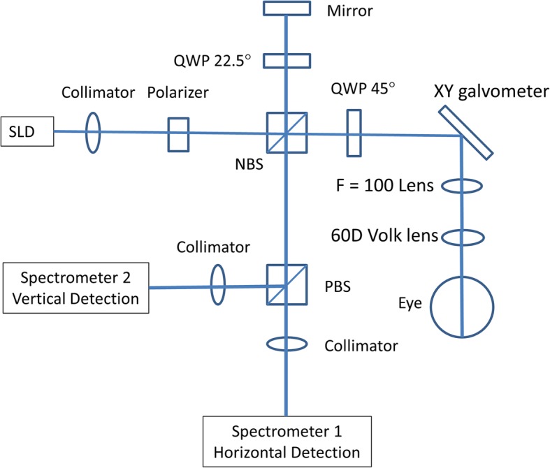
The retina has been used to study the pathophysiology of multiple sclerosis (MS). Peripapillary retinal nerve fiber layer (pRNFL) thinning has been suggested as an ocular biomarker of neurodegeneration in MS. The goal of this project was to determine the birefringence of the pRNFL by measuring the fiber birefringence using polarization sensitive optical coherence tomography (PS-OCT).

METHODS

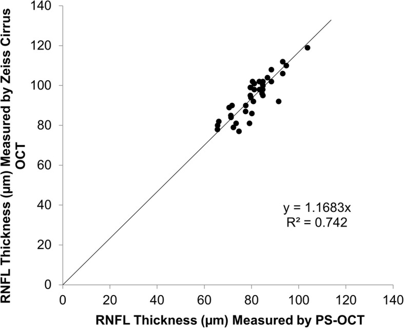
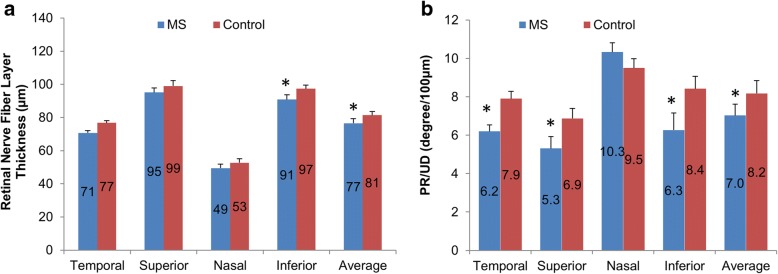
Sixty-six MS patients without history of optic neuritis (age: 39.9 ± 11.0 yrs. old, 53 females and 13 males) and 66 age- and gender-matched normal controls (age: 40.7 ± 11.4 yrs. old) were recruited. Custom built PS-OCT was used to measure phase retardation per unit depth (PR/UD, proportional to the birefringence) and pRNFL thickness in each quadrant of the pRNFL. In addition, clinical manifestation was used to correlate with the pRNFL birefringence.

RESULTS

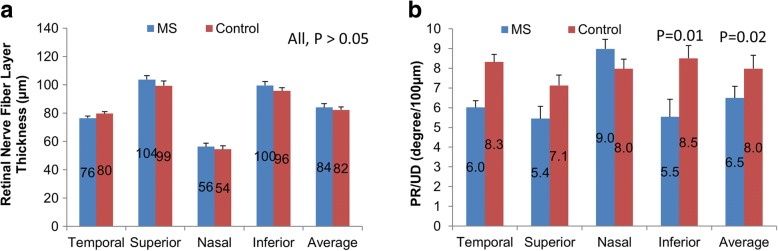
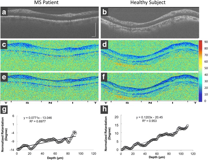
The pRNFL was thinner in the temporal and inferior quadrants in MS patients compared with normal controls ( < 0.05). The PR/UD of the pRNFL was significantly decreased in MS patients (P < 0.05) in all quadrants except for the nasal quadrant. In both groups, the PR/UD from all four quadrants was not related to the averaged pRNFL thickness ( > 0.05). In MS patients, the PR/UD was not related to the expanded disability status scale (EDSS) nor disease duration (r ranged from - 0.17 to 0.02,  > 0.05).

CONCLUSION

This is the first study using PS-OCT to study the pRNFL birefringence in MS patients. Decreased birefringence of the pRNFL may indicate microtubule abnormality, and could be a potential biomarker for detecting early neurodegeneration in MS.

摘要

背景

视网膜已被用于研究多发性硬化症(MS)的病理生理学。视乳头周围视网膜神经纤维层(pRNFL)变薄被认为是MS神经退行性变的一种眼部生物标志物。本项目的目的是通过使用偏振敏感光学相干断层扫描(PS-OCT)测量纤维双折射来确定pRNFL的双折射。

方法

招募了66例无视神经炎病史的MS患者(年龄:39.9±11.0岁,53例女性和13例男性)以及66例年龄和性别匹配的正常对照(年龄:40.7±11.4岁)。使用定制的PS-OCT测量pRNFL每个象限的单位深度相位延迟(PR/UD,与双折射成正比)和pRNFL厚度。此外,将临床表现与pRNFL双折射进行关联。

结果

与正常对照相比,MS患者颞侧和下方象限的pRNFL更薄(P<0.05)。除鼻侧象限外,MS患者所有象限的pRNFL的PR/UD均显著降低(P<0.05)。在两组中,所有四个象限的PR/UD均与平均pRNFL厚度无关(P>0.05)。在MS患者中,PR/UD与扩展残疾状态量表(EDSS)和病程均无关(r范围为-0.17至0.02,P>0.05)。

结论

这是第一项使用PS-OCT研究MS患者pRNFL双折射的研究。pRNFL双折射降低可能表明微管异常,并且可能是检测MS早期神经退行性变的潜在生物标志物。

https://cdn.ncbi.nlm.nih.gov/pmc/blobs/a22c/6011597/5d547db4b97b/40662_2018_108_Fig5_HTML.jpg
https://cdn.ncbi.nlm.nih.gov/pmc/blobs/a22c/6011597/6a1a9d1edf5c/40662_2018_108_Fig1_HTML.jpg
https://cdn.ncbi.nlm.nih.gov/pmc/blobs/a22c/6011597/ca59adb0c596/40662_2018_108_Fig2_HTML.jpg
https://cdn.ncbi.nlm.nih.gov/pmc/blobs/a22c/6011597/1d70297a4e3d/40662_2018_108_Fig3_HTML.jpg
https://cdn.ncbi.nlm.nih.gov/pmc/blobs/a22c/6011597/233c254d5578/40662_2018_108_Fig4_HTML.jpg
https://cdn.ncbi.nlm.nih.gov/pmc/blobs/a22c/6011597/5d547db4b97b/40662_2018_108_Fig5_HTML.jpg

相似文献

1
Altered birefringence of peripapillary retinal nerve fiber layer in multiple sclerosis measured by polarization sensitive optical coherence tomography.利用偏振敏感光学相干断层扫描技术测量多发性硬化症患者视乳头周围视网膜神经纤维层的双折射改变
Eye Vis (Lond). 2018 Jun 17;5:14. doi: 10.1186/s40662-018-0108-z. eCollection 2018.
2
Retinal nerve fiber layer thickness associates with cognitive impairment and physical disability in multiple sclerosis.视网膜神经纤维层厚度与多发性硬化症患者的认知障碍和身体残疾有关。
Mult Scler Relat Disord. 2019 Nov;36:101414. doi: 10.1016/j.msard.2019.101414. Epub 2019 Sep 26.
3
OCT retinal nerve fiber layer thickness differentiates acute optic neuritis from MOG antibody-associated disease and Multiple Sclerosis: RNFL thickening in acute optic neuritis from MOGAD vs MS.光学相干断层扫描(OCT)测量的视网膜神经纤维层厚度可区分急性视神经炎与MOG抗体相关疾病及多发性硬化症:MOG抗体相关疾病与多发性硬化症所致急性视神经炎的视网膜神经纤维层增厚情况比较
Mult Scler Relat Disord. 2022 Feb;58:103525. doi: 10.1016/j.msard.2022.103525. Epub 2022 Jan 11.
4
Birefringence of the primate retinal nerve fiber layer.灵长类视网膜神经纤维层的双折射
Exp Eye Res. 2005 Jul;81(1):81-9. doi: 10.1016/j.exer.2005.01.009.
5
Retinal thickness measured with optical coherence tomography and risk of disability worsening in multiple sclerosis: a cohort study.光学相干断层扫描测量的视网膜厚度与多发性硬化症残疾恶化风险:一项队列研究。
Lancet Neurol. 2016 May;15(6):574-84. doi: 10.1016/S1474-4422(16)00068-5. Epub 2016 Mar 18.
6
Optical Coherence Tomography in Neuromyelitis Optica spectrum disorder and Multiple Sclerosis: A population-based study.视神经脊髓炎谱系疾病和多发性硬化症的光学相干断层扫描:一项基于人群的研究。
Mult Scler Relat Disord. 2021 Jan;47:102625. doi: 10.1016/j.msard.2020.102625. Epub 2020 Nov 14.
7
Peripapillary retinal nerve fibre layer as measured by optical coherence tomography is a prognostic biomarker not only for physical but also for cognitive disability progression in multiple sclerosis.光学相干断层扫描测量的视盘周围视网膜神经纤维层不仅是多发性硬化症身体残疾进展的预后生物标志物,也是认知残疾进展的预后生物标志物。
Mult Scler. 2019 Feb;25(2):196-203. doi: 10.1177/1352458517740216. Epub 2017 Nov 2.
8
Retinal Nerve Fiber Layer Thickness Correlates with Serum and Cerebrospinal Fluid Neurofilament Levels and is Associated with Current Disability in Multiple Sclerosis.视网膜神经纤维层厚度与血清及脑脊液神经丝水平相关,并与多发性硬化症患者当前的残疾状况有关。
Noro Psikiyatr Ars. 2021 Jan 16;58(1):34-40. doi: 10.29399/npa.27355. eCollection 2021 Mar.
9
Retina ganglion cell/inner plexiform layer and peripapillary nerve fiber layer thickness in patients with acromegaly.肢端肥大症患者的视网膜神经节细胞/内网状层及视乳头周围神经纤维层厚度
Int Ophthalmol. 2017 Jun;37(3):591-598. doi: 10.1007/s10792-016-0310-8. Epub 2016 Aug 4.
10
Clinical effects associated with five-year retinal nerve fiber layer thinning in multiple sclerosis.多发性硬化症五年视网膜神经纤维层变薄的临床影响。
J Neurol Sci. 2021 Aug 15;427:117552. doi: 10.1016/j.jns.2021.117552. Epub 2021 Jun 23.

引用本文的文献

1
Single-input polarization-sensitive optical coherence tomography through a catheter.通过导管的单输入偏振敏感光学相干断层扫描术
Biomed Opt Express. 2023 Aug 10;14(9):4609-4626. doi: 10.1364/BOE.497123. eCollection 2023 Sep 1.
2
Machine learning classification of multiple sclerosis in children using optical coherence tomography.基于光学相干断层扫描的机器学习在儿童多发性硬化症中的分类。
Mult Scler. 2022 Dec;28(14):2253-2262. doi: 10.1177/13524585221112605. Epub 2022 Aug 9.
3
Decreased Vessel Density in Retinal Capillary Plexus and Thinner Ganglion Cell Complex Associated With Cognitive Impairment.

本文引用的文献

1
Retinal layer segmentation in multiple sclerosis: a systematic review and meta-analysis.多发性硬化症中的视网膜层分割:系统评价和荟萃分析。
Lancet Neurol. 2017 Oct;16(10):797-812. doi: 10.1016/S1474-4422(17)30278-8. Epub 2017 Sep 12.
2
Retinal Nerve Fiber Layer Thickness Measurement Comparison Using Spectral Domain and Swept Source Optical Coherence Tomography.使用光谱域和扫频光学相干断层扫描技术比较视网膜神经纤维层厚度测量
Korean J Ophthalmol. 2016 Apr;30(2):140-7. doi: 10.3341/kjo.2016.30.2.140. Epub 2016 Mar 25.
3
Retinal nerve fiber layer thickness, brain atrophy, and disability in multiple sclerosis patients.
视网膜毛细血管丛中血管密度降低以及神经节细胞复合体变薄与认知障碍相关。
Front Aging Neurosci. 2022 Apr 26;14:872466. doi: 10.3389/fnagi.2022.872466. eCollection 2022.
4
Focal Loss Analysis of Nerve Fiber Layer Reflectance for Glaucoma Diagnosis.用于青光眼诊断的神经纤维层反射的焦点损失分析。
Transl Vis Sci Technol. 2021 May 3;10(6):9. doi: 10.1167/tvst.10.6.9.
5
Longitudinal Study of Retinal Structure, Vascular, and Neuronal Function in Patients With Relapsing-Remitting Multiple Sclerosis: 1-Year Follow-Up.复发缓解型多发性硬化症患者的视网膜结构、血管和神经元功能的纵向研究:1 年随访。
Transl Vis Sci Technol. 2021 May 3;10(6):6. doi: 10.1167/tvst.10.6.6.
6
Identification of Human Pathological Mitral Chordae Tendineae Using Polarization-sensitive Optical Coherence Tomography.采用偏振敏感光学相干断层成像术鉴定人类病理性二尖瓣腱索。
Sensors (Basel). 2019 Jan 28;19(3):543. doi: 10.3390/s19030543.
7
Retinal nerve fiber layer (RNFL) integrity and its relations to retinal microvasculature and microcirculation in myopic eyes.近视眼中视网膜神经纤维层(RNFL)的完整性及其与视网膜微血管和微循环的关系。
Eye Vis (Lond). 2018 Oct 10;5:25. doi: 10.1186/s40662-018-0120-3. eCollection 2018.
多发性硬化症患者的视网膜神经纤维层厚度、脑萎缩与残疾。
J Neuroophthalmol. 2014 Mar;34(1):23-8. doi: 10.1097/WNO.0000000000000057.
4
Comparative assessment for the ability of Cirrus, RTVue, and 3D-OCT to diagnose glaucoma.比较评估 Cirrus、RTVue 和 3D-OCT 诊断青光眼的能力。
Invest Ophthalmol Vis Sci. 2013 Jul 10;54(7):4478-84. doi: 10.1167/iovs.12-11268.
5
DTI Measurements in Multiple Sclerosis: Evaluation of Brain Damage and Clinical Implications.多发性硬化症中的扩散张量成像测量:脑损伤评估及临床意义
Mult Scler Int. 2013;2013:671730. doi: 10.1155/2013/671730. Epub 2013 Mar 31.
6
Measuring retinal nerve fiber layer birefringence, retardation, and thickness using wide-field, high-speed polarization sensitive spectral domain OCT.利用宽视野、高速偏振敏感光谱域 OCT 测量视网膜神经纤维层双折射、延迟和厚度。
Invest Ophthalmol Vis Sci. 2013 Jan 7;54(1):72-84. doi: 10.1167/iovs.12-10089.
7
Reflectance decreases before thickness changes in the retinal nerve fiber layer in glaucomatous retinas.在青光眼视网膜中,神经纤维层的厚度发生变化之前,反射率会降低。
Invest Ophthalmol Vis Sci. 2011 Aug 24;52(9):6737-42. doi: 10.1167/iovs.11-7665.
8
Polarization sensitive optical coherence tomography in the human eye.偏振敏感光学相干断层扫描在人眼中的应用。
Prog Retin Eye Res. 2011 Nov;30(6):431-51. doi: 10.1016/j.preteyeres.2011.06.003. Epub 2011 Jun 26.
9
The relationship between retinal ganglion cell axon constituents and retinal nerve fiber layer birefringence in the primate.灵长类动物视网膜神经节细胞轴突成分与视网膜神经纤维层双折射之间的关系。
Invest Ophthalmol Vis Sci. 2009 Nov;50(11):5238-46. doi: 10.1167/iovs.08-3263. Epub 2009 Jun 3.
10
Measurement and imaging of birefringence and optic axis orientation by phase resolved polarization sensitive optical coherence tomography.通过相位分辨偏振敏感光学相干断层扫描技术测量双折射和光轴方向并进行成像。
Opt Express. 2001 Dec 17;9(13):780-90. doi: 10.1364/oe.9.000780.