Sbardella Emilia, Tona Francesca, Petsas Nikolaos, Pantano Patrizia
Department of Neurology and Psychiatry, Sapienza University, Viale dell'Università 30, 00185 Rome, Italy.
Mult Scler Int. 2013;2013:671730. doi: 10.1155/2013/671730. Epub 2013 Mar 31.
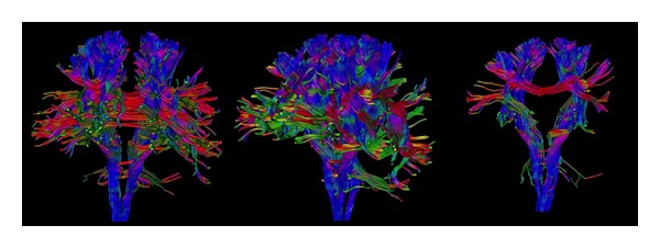
Diffusion tensor imaging (DTI) is an effective means of quantifying parameters of demyelination and axonal loss. The application of DTI in Multiple Sclerosis (MS) has yielded noteworthy results. DTI abnormalities, which are already detectable in patients with clinically isolated syndrome (CIS), become more pronounced as disease duration and neurological impairment increase. The assessment of the microstructural alterations of white and grey matter in MS may shed light on mechanisms responsible for irreversible disability accumulation. In this paper, we examine the DTI analysis methods, the results obtained in the various tissues of the central nervous system, and correlations with clinical features and other MRI parameters. The adoption of DTI metrics to assess the outcome of prognostic measures may represent an extremely important step forward in the MS research field.
扩散张量成像(DTI)是量化脱髓鞘和轴突损失参数的有效手段。DTI在多发性硬化症(MS)中的应用已取得显著成果。DTI异常在临床孤立综合征(CIS)患者中已可检测到,随着疾病持续时间和神经功能障碍的增加而变得更加明显。对MS中白质和灰质微观结构改变的评估可能有助于揭示不可逆残疾积累的机制。在本文中,我们研究了DTI分析方法、在中枢神经系统各组织中获得的结果,以及与临床特征和其他MRI参数的相关性。采用DTI指标评估预后措施的结果可能是MS研究领域向前迈出的极其重要的一步。