Song Woo Keun, Cho Ah Ran, Yoon Young Hee
Department of Ophthalmology, College of Medicine, University of Ulsan, Asan Medical Center, 88 Olympic-ro 43-Gil, Songpa-gu, Seoul, 05505, South Korea.
BMC Ophthalmol. 2018 Jun 28;18(1):156. doi: 10.1186/s12886-018-0832-0.
To describe a case of highly suspected primary intraocular lymphoma (PIOL) in a patient using etanercept for the treatment of rheumatoid arthritis.
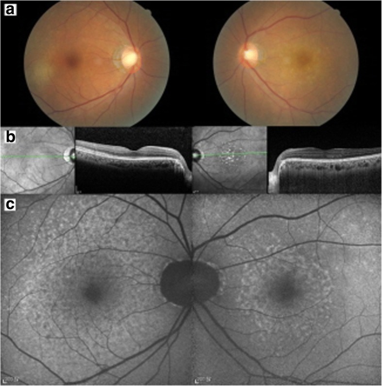
A 50-year-old female patient presented with decreased vision in her left eye that lasted for a week. She had a 15-year history of seropositive rheumatoid arthritis (RA), and had been taking weekly etanercept for the preceding 8 months. Funduscopic examination and SD-OCT showed a swollen ellipsoid zone (EZ) and a retinal pigment epithelium (RPE) irregularity of the right eye. We also noted EZ disruption and a RPE irregularity in the left eye. As subretinal infiltration was aggravated in the right eye after the initial treatment, we completed a vitrectomy. Vitreous cytology revealed PIOL with positive CD20 immunostaining. She was treated with serial intravitreal methotrexate injections and systemic chemotherapy. After the treatment, subretinal infiltration and subRPE deposits were decreased in the right eye with no evidence of recurrence in either eye.
This case suggests a potential relationship between immunosuppression with anti-TNFα medication, and increased risk for lymphoma, especially in patients with underlying rheumatologic disorders and especially in patients with suspected chronic refractory uveitis.
描述一例使用依那西普治疗类风湿关节炎的患者高度疑似原发性眼内淋巴瘤(PIOL)的病例。
一名50岁女性患者出现左眼视力下降,持续一周。她有15年血清阳性类风湿关节炎(RA)病史,在过去8个月中每周使用依那西普治疗。眼底检查和频域光学相干断层扫描(SD - OCT)显示右眼椭圆体带(EZ)肿胀和视网膜色素上皮(RPE)不规则。我们还注意到左眼EZ破坏和RPE不规则。由于初始治疗后右眼视网膜下浸润加重,我们完成了玻璃体切除术。玻璃体细胞学检查显示PIOL,CD20免疫染色呈阳性。她接受了系列玻璃体内甲氨蝶呤注射和全身化疗。治疗后,右眼视网膜下浸润和RPE下沉积物减少,双眼均无复发迹象。
本病例提示抗TNFα药物免疫抑制与淋巴瘤风险增加之间可能存在关联,尤其是在患有潜在风湿性疾病的患者中,特别是在疑似慢性难治性葡萄膜炎的患者中。