Cardiovascular Intervention, St. Carollo Hospital, Suncheon-si, Jeollanam-do, South Korea.
Department of Internal Medicine, St. Carollo Hospital, Suncheon-si, Jeollanam-do, South Korea.
Cardiovasc Drugs Ther. 2018 Aug;32(4):329-338. doi: 10.1007/s10557-018-6804-z.
BACKGROUND/AIMS: The progression and development of congestive heart failure is still considered a large problem despite the existence of revascularization therapies and optimal, state-of-the-art medical services. An acute myocardial infarction (AMI) is a major cause of congestive heart failure, so researchers are investigating techniques to complement primary percutaneous coronary intervention (PCI) or thrombolytic therapy to prevent congestive heart failure after AMI.
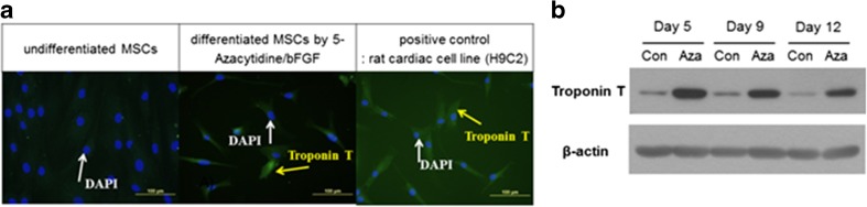
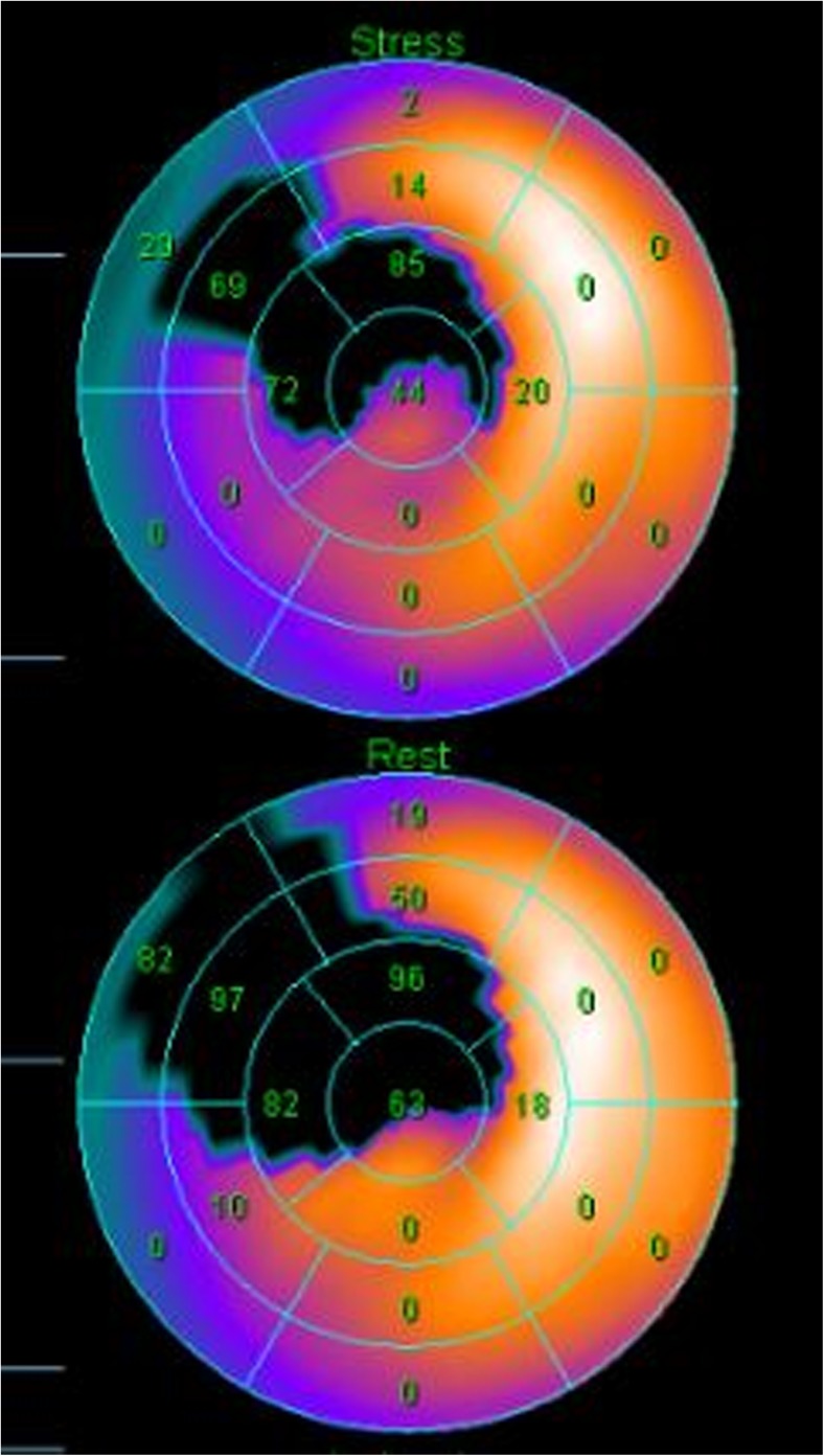
Twenty-six patients with successful PCI for acute ST-segment elevation anterior wall myocardial infarction were assigned to either a control group (n = 12) or a bone marrow mesenchymal stem cells (BM-MSC) group (n = 14). The control group received optimum post-infarction treatment, and the BMSC group received intracoronary delivery of autologous BMSC at 1 month after PCI with the optimum medical treatment. The primary endpoint was a left ventricular ejection fraction (LVEF) change from baseline to 4-month follow-up, as determined via myocardial single-photon emission computed tomography (SPECT).
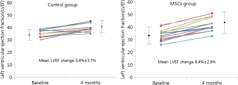
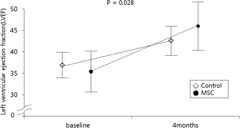
The global LVEF at baseline (determined 3.5 ± 1.5 days after PCI) was 35.4 ± 3.0% in the control group and 33.6 ± 4.7% in the BM-MSC group. BMSC transfer enhanced left ventricular systolic function primarily in anterior wall myocardial segments adjacent to the LAD infarcted area. Four months later, via SPECT, global LVEF had increased by 4.8 ± 1.9% in the control group and 8.8 ± 2.9% in the BM-MSC group (p = 0.031). The cell transfer did not increase the risk of adverse clinical events, in-stent restenosis, or proarrhythmic effects. The echocardiographic evaluation also revealed a significant increase in the LVEF value from baseline to the 4-month (9.0 ± 4.7 and 5.3 ± 2.6%, p = 0.023) and 12-month (9.9 ± 5.2% and 6.5 ± 2.7%, p = 0.048) follow-up in the BM-MSC group but not in the control group.
Intracoronary administration of autologous BM-MSC was tolerable and safe with significant improvement in LVEF at 4-month (SPECT and echocardiography result) and 12-month (echocardiography result only) follow-up in patients with anterior AMI.
背景/目的:尽管存在血运重建治疗和最佳的现代医疗服务,充血性心力衰竭的进展和发展仍然被认为是一个大问题。急性心肌梗死(AMI)是充血性心力衰竭的主要原因,因此研究人员正在研究技术,以补充经皮冠状动脉介入治疗(PCI)或溶栓治疗,以防止 AMI 后发生充血性心力衰竭。
26 例急性 ST 段抬高前壁心肌梗死患者行 PCI 成功,分为对照组(n=12)和骨髓间充质干细胞(BM-MSC)组(n=14)。对照组接受最佳梗死后治疗,BM-MSC 组在 PCI 后 1 个月接受最佳药物治疗的自体 BM-MSC 冠状动脉内给药。主要终点是通过心肌单光子发射计算机断层扫描(SPECT)从基线到 4 个月随访时左心室射血分数(LVEF)的变化。
对照组(PCI 后 3.5±1.5 天)的基线时整体 LVEF 为 35.4±3.0%,BM-MSC 组为 33.6±4.7%。BM-MSC 转移主要增强了与 LAD 梗死区域相邻的前壁心肌节段的左心室收缩功能。4 个月后,通过 SPECT,对照组的整体 LVEF 增加了 4.8±1.9%,BM-MSC 组增加了 8.8±2.9%(p=0.031)。细胞转移并未增加不良临床事件、支架内再狭窄或致心律失常作用的风险。超声心动图评估还显示,从基线到 4 个月(9.0±4.7 和 5.3±2.6%,p=0.023)和 12 个月(9.9±5.2%和 6.5±2.7%,p=0.048)随访时,BM-MSC 组的 LVEF 值显著增加,但对照组无明显变化。
自体 BM-MSC 冠状动脉内给药是安全耐受的,在前壁 AMI 患者中,4 个月(SPECT 和超声心动图结果)和 12 个月(仅超声心动图结果)随访时 LVEF 显著改善。