Abd-El Khalek Abd-ALRazek Ahmed, Fahmy Dalia Monir
J Comput Assist Tomogr. 2018 Sep/Oct;42(5):688-696. doi: 10.1097/RCT.0000000000000754.
The objective of this study is to evaluate the role of diffusion-weighted imaging (DWI) in assessment of the activity of Crohn disease (CD) and to explore differences between DWI in 3 T and 1.5 T.
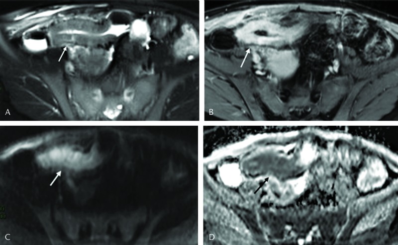
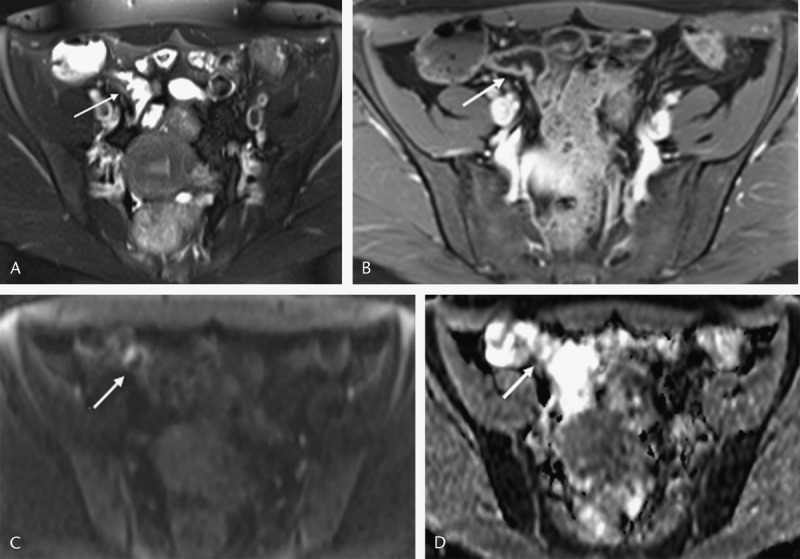
Postcontrast magnetic resonance enterography with DWI of 72 patients with pathological proof of CD was retrospectively evaluated for restricted diffusion qualitatively and quantitavely in 3 T (n = 40) and 1.5 T (n = 32). Magnetic resonance activity score of 7 or higher was used as reference of activity.
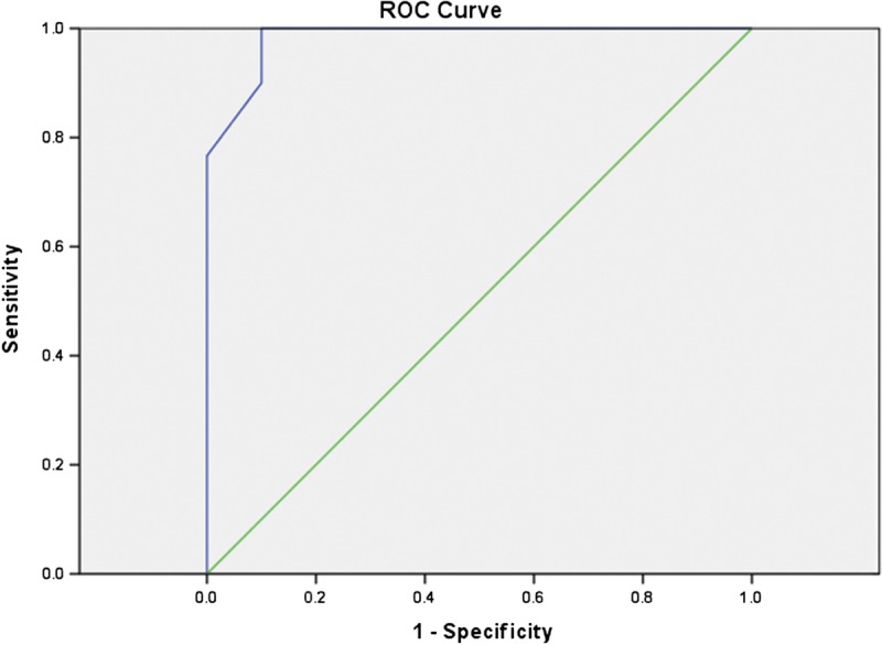
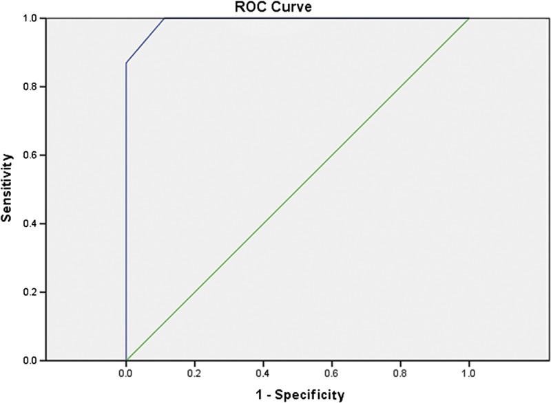
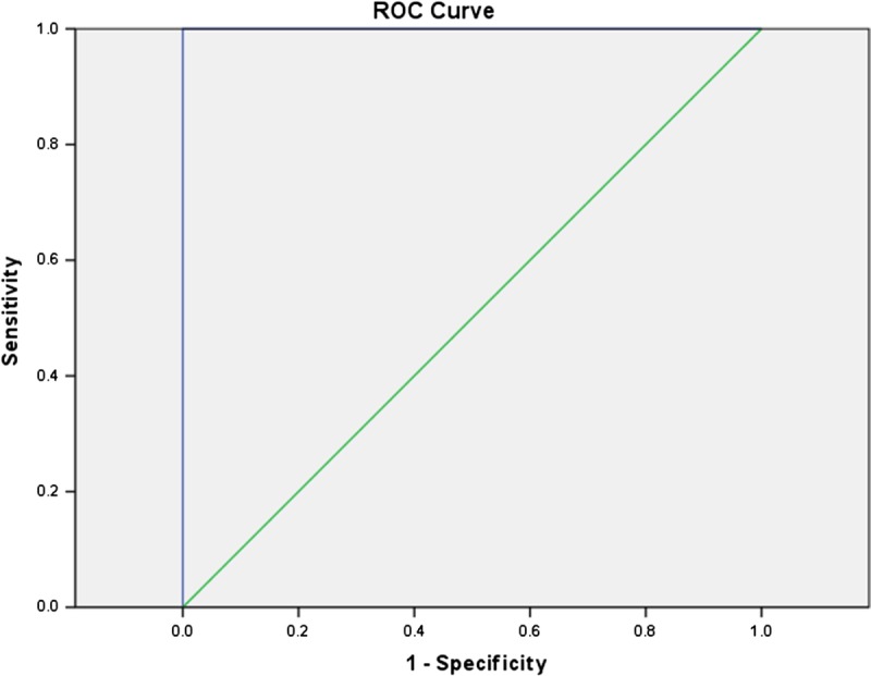
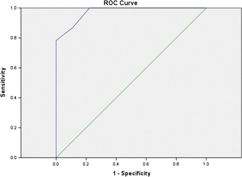
Fifty-five patients had active lesions. Diffusion-weighted imaging hyperintensity showed sensitivity (100%, 100%) and specificity (88.89%, 100%) in 1.5/3 T for activity assessment. Mean ± SD apparent diffusion coefficient for active lesions was 1.21 ± 0.42 and 1.28 ± 0.59 × 10 mm/s in 1.5 and 3 T, respectively. The proposed cutoff values of 1.35 and 1.38 × 10 mm/s in 1.5 and 3 T, respectively, had sensitivity (80%, 93%), specificity (100%, 90%), accuracy (88%, 93%), and no significant difference in accuracy between 1.5/3 T (P = 0.48).
Diffusion-weighted imaging hypersensitivity and apparent diffusion coefficient values accurately assessed the activity of CD. No significant statistical difference in diagnostic accuracy was detected between 1.5 and 3 T.
本研究旨在评估扩散加权成像(DWI)在评估克罗恩病(CD)活动度中的作用,并探讨3T和1.5T条件下DWI的差异。
回顾性评估72例经病理证实为CD患者的增强磁共振肠造影及DWI图像,分别在3T(n = 40)和1.5T(n = 32)条件下对扩散受限进行定性和定量分析。以磁共振活动评分7分及以上作为活动度参考标准。
55例患者有活动性病变。DWI高信号在1.5T/3T条件下评估活动度的敏感度为(100%,100%),特异度为(88.89%,100%)。1.5T和3T条件下活动性病变的平均表观扩散系数(Mean ± SD)分别为1.21 ± 0.42和1.28 ± 0.59×10⁻³mm²/s。1.5T和3T条件下分别设定的表观扩散系数截断值1.35×10⁻³mm²/s和1.38×10⁻³mm²/s,敏感度分别为(80%,93%),特异度分别为(100%,90%),准确度分别为(88%,93%),1.5T/3T之间准确度无显著差异(P = 0.48)。
DWI高敏感度及表观扩散系数值可准确评估CD的活动度。1.5T和3T条件下诊断准确度无显著统计学差异。