Ayed Mohammed S, Shafiq Sami S, Diab Hafez M, Alahmari Ahmed D, Divakar Darshan D
Aseer Region, Abha, Kingdom of Saudi Arabia. E-mail.
Saudi Med J. 2018 Jul;39(7):719-724. doi: 10.15537/smj.2018.7.22453.
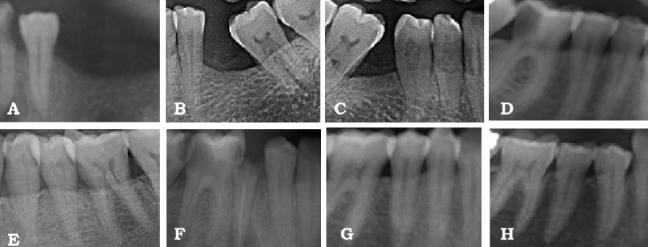
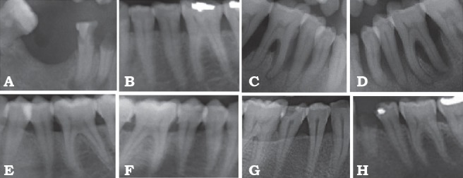
To assess the usefulness of periapical dental radiograph as a screening tool aimed at early signs of osteoporosis in postmenopausal periodontal patients and root surface evaluation using spectrochemical analysis. Methods: This study was conducted at the Department of Periodontics, Riyadh Elm University, Riyadh, Saudi Arabia, for 12 months between December 2016 and November 2017. Two groups consisted healthy postmenopausal women having chronic periodontitis and postmenopausal women having chronic periodontitis with osteoporosis. Osteoporosis were evaluated for plaque index (PI); gingival index (GI); clinical attachment level (CAL); probing pocket depth (PPD), and bone mineral density (BMD). A standardized digital dental periapical radiographs were taken for every patient. The spectrochemical analysis was carried out using the self-assembled Laser-Induced Breakdown Spectroscopy (LIBS) system used for qualitative and quantitative analysis of Calcium (Ca), Potassium (K), Phosphorus (P), Fluoride (F), and Magnesium (Mg). Results: There was no statistically significant difference between both groups for GI and PI. Similarly PPD and CAL were showing the difference but statistically, significant difference was only for CAL. Value of distance starting from cement enamel junction to the alveolar crest (CEJ-AC) and BMD were having a statistically significant variance between both groups. The differences between osteoporotic and control group were statistically significant regarding Ca with the mean higher in the control group. Furthermore, the variances between the groups in both K and Mg were statistically significant with higher mean in the osteoporotic group (p less than 0.05). Conclusion: The clinical, radiographic, and experimental findings of this study indicated that osteoporosis has a direct effect on the progression rate of periodontal tissue destruction and dental radiographic can be suggested as a screening tool for an early sign of osteoporosis.
评估根尖牙片作为绝经后牙周病患者骨质疏松早期迹象筛查工具的实用性以及使用光谱化学分析进行根面评估。方法:本研究于2016年12月至2017年11月在沙特阿拉伯利雅得伊玛目大学牙周病科进行,为期12个月。两组分别为患有慢性牙周炎的健康绝经后女性和患有慢性牙周炎伴骨质疏松的绝经后女性。评估骨质疏松情况的指标包括菌斑指数(PI)、牙龈指数(GI)、临床附着水平(CAL)、探诊深度(PPD)和骨密度(BMD)。为每位患者拍摄标准化的数字化根尖牙片。使用自行组装的激光诱导击穿光谱(LIBS)系统进行光谱化学分析,用于对钙(Ca)、钾(K)、磷(P)、氟(F)和镁(Mg)进行定性和定量分析。结果:两组在GI和PI方面无统计学显著差异。同样,PPD和CAL存在差异,但在统计学上,仅CAL有显著差异。从牙骨质釉质界到牙槽嵴的距离(CEJ - AC)值和BMD在两组之间存在统计学显著差异。骨质疏松组和对照组在Ca方面的差异具有统计学意义,对照组的平均值更高。此外,两组在K和Mg方面的差异也具有统计学意义,骨质疏松组的平均值更高(p小于0.05)。结论:本研究的临床、影像学和实验结果表明,骨质疏松对牙周组织破坏的进展速度有直接影响,根尖牙片可作为骨质疏松早期迹象的筛查工具。