Paulsson Johan O, Zedenius Jan, Juhlin C Christofer
Department of Oncology-Pathology, Karolinska Institutet, Stockholm, Sweden.
Department of Molecular Medicine and Surgery, Karolinska Institutet, Stockholm, Sweden.
BMC Endocr Disord. 2018 Jul 5;18(1):46. doi: 10.1186/s12902-018-0275-x.
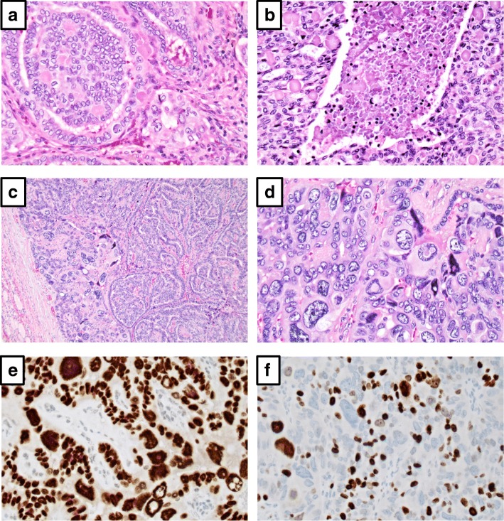
Papillary thyroid carcinoma with pleomorphic tumor giant cells (PTC-PC) is characterized by the occurrence of bizarre, pleomorphic cells within a small area of a conventional PTC. The histologic distinction between PTC-PC and PTC's with a focal anaplastic thyroid cancer (ATC) component (denoted in the 2004 WHO classification as "papillary thyroid carcinoma with spindle and giant cell carcinoma", PTC-SGC) is debated, however the prognosis is thought to be different (excellent for PTC-PC, poor for PTC-SGC). Therefore, this diagnostic challenge is significant for any endocrine pathologist to recognize. Herein, we report the histological and clinical workup of a PTC-PC case, with particular focus on the molecular analyses that facilitated the establishment of the final diagnosis.
The patient was a pregnant, 28-year-old female presenting with a 30 mm conventional PTC, with focal areas with undifferentiated cells exhibiting exaggerated nuclear pleomorphism. No foci of extrathyroidal extension, angioinvasion or lymph node engagement were seen. Immunohistochemical analyses revealed the pleomorphic cells exhibiting retained differentiation. Molecular genetic analyses demonstrated a codon V600 missense mutation of the BRAF gene, but no TP53 or TERT promoter mutations. The absence of an aggressive phenotype in addition to the lack of mutations in two major ATC-related genes led to the diagnosis of a PTC-PC. Postoperative MRI showed no evidence of metastatic disease. Radioiodine ablation was performed seven months post-operatively, and a SPECT-CT imaging did not show signs of residual tissue. She is well and without signs of disease 16 months post-operatively.
PTC-PC is a differential diagnosis to PTC-SGC that mandates careful considerations. Taken together with previous publications, PTC-PC seems to be histologically similar to PTC-SGC, but clinically distinct. Even so, the distinction is not easily made given the different therapeutic consequences for each individual patient. This is the first report that includes molecular genetics to aid in finalizing the diagnosis. Exclusion of mutations in TP53 and the TERT promoter could be considered as an adjunct tool when assessing papillary thyroid cancer with focal pleomorphism.
伴有多形性肿瘤巨细胞的甲状腺乳头状癌(PTC-PC)的特征是在传统PTC的小区域内出现奇异的多形性细胞。PTC-PC与具有局灶性间变性甲状腺癌(ATC)成分的PTC(在2004年WHO分类中称为“伴有梭形和巨细胞癌的甲状腺乳头状癌”,PTC-SGC)之间的组织学区分存在争议,然而其预后被认为有所不同(PTC-PC预后良好,PTC-SGC预后不良)。因此,对于任何内分泌病理学家来说,认识到这一诊断挑战都很重要。在此,我们报告一例PTC-PC病例的组织学和临床检查,特别关注有助于最终诊断确立的分子分析。
患者为一名28岁的孕妇,患有一个30mm的传统PTC,局部区域有未分化细胞,表现出核多形性加剧。未见甲状腺外扩展、血管侵犯或淋巴结受累灶。免疫组织化学分析显示多形性细胞保留了分化特征。分子遗传学分析显示BRAF基因密码子V600错义突变,但无TP53或TERT启动子突变。除了两个主要的ATC相关基因无突变外,缺乏侵袭性表型,从而诊断为PTC-PC。术后MRI未显示转移疾病的证据。术后七个月进行了放射性碘消融,SPECT-CT成像未显示残留组织迹象。术后16个月,她情况良好,无疾病迹象。
PTC-PC是PTC-SGC的鉴别诊断,需要仔细考虑。结合先前的文献,PTC-PC在组织学上似乎与PTC-SGC相似,但临床特征不同。即便如此,鉴于每个患者的治疗后果不同,这种区分并不容易做出。这是第一份包括分子遗传学以辅助最终诊断的报告。在评估具有局灶性多形性的甲状腺乳头状癌时,排除TP53和TERT启动子突变可被视为一种辅助工具。