Zhuang Nan, Zhu Qingli, Li Wenbo, Wang Miaoqian, Yang Qian, Liu Wei, Li Ji, Yang Hong, Zhou Weixun
Department of Ultrasound Department of Radiology Department of Gastroenterology Department of Pathology, Peking Union Medical College Hospital, Beijing, China.
Medicine (Baltimore). 2018 Jul;97(27):e11407. doi: 10.1097/MD.0000000000011407.
Primary lymphoma that arises from the intestine is an uncommon malignant tumour, while intestinal fistula caused by primary lymphoma is even rarer. Non-specific clinical performance makes early diagnosis difficult, although imaging modalities might play an essential role in the detection of intestinal fistula.
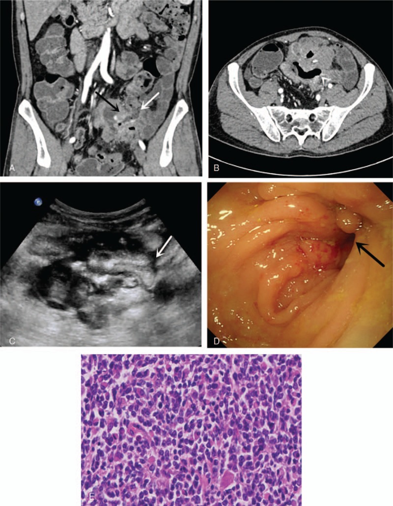
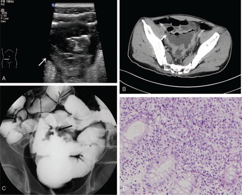
Patient 1: A 60-year-old male hospitalized with diarrhoea and abdominal pain for seven months underwent computed tomography enterography (CTE) that demonstrated ileum internal fistula and ileac-sigmoid colon fistula. Ultrasound (US) showed small intestinal wall thickened and development of a fistula of the sigmoid colon due to malignance. Patient 2: A 43-year-old male presented with abdominal pain and diarrhoea lasting one year. US revealed a fistula between the sigmoid colon and the ileum, and CTE showed that the wall of the partial sigmoid colon was abnormally thickened and enhanced with an ileal-sigmoid fistula that strongly suggested the diagnosis of lymphoma.
Both the two patients were diagnosed as intestinal fistula caused by primary non-Hodgkin's intestinal lymphoma.
The patient 1 underwent surgery followed by chemotherapy. The patient 2 accepted chemotherapy.
Two patients' general conditions remained stable and the imaging revealed no recurrence after follow-up of about 12 months.
Cross-sectional imaging, such as US and CT, plays an essential role in intestinal lymphoma fistula diagnosis.
原发性肠道淋巴瘤是一种罕见的恶性肿瘤,而由原发性淋巴瘤引起的肠瘘则更为罕见。尽管影像学检查在肠瘘的检测中可能起着至关重要的作用,但非特异性的临床表现使得早期诊断困难。
患者1:一名60岁男性因腹泻和腹痛住院7个月,接受计算机断层扫描小肠造影(CTE)检查,显示回肠内瘘和回肠-乙状结肠瘘。超声(US)显示小肠壁增厚,因恶性肿瘤导致乙状结肠瘘形成。患者2:一名43岁男性出现腹痛和腹泻持续1年。超声显示乙状结肠和回肠之间有瘘管,CTE显示部分乙状结肠壁异常增厚并强化,伴有回肠-乙状结肠瘘,强烈提示淋巴瘤诊断。
两名患者均被诊断为原发性非霍奇金肠道淋巴瘤所致肠瘘。
患者1接受手术,随后进行化疗。患者2接受化疗。
两名患者的一般情况保持稳定,随访约12个月后影像学检查未显示复发。
超声和CT等横断面成像在肠道淋巴瘤瘘的诊断中起着至关重要的作用。