District Hospital, Cardiology, Radom, Poland.
Sonovum AG, Leipzig, Germany.
PLoS One. 2018 Jul 6;13(7):e0199999. doi: 10.1371/journal.pone.0199999. eCollection 2018.
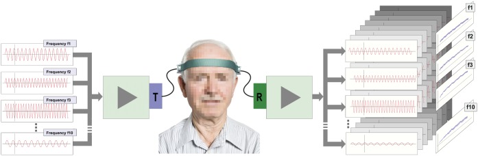
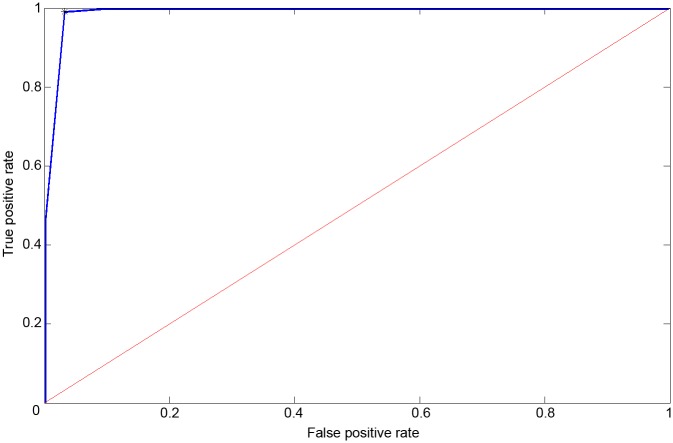
Acoustocerebrography is a novel, non-invasive, transcranial ultrasonic diagnostic method based on the transmission of multispectral ultrasound signals propagating through the brain tissue. Dedicated signal processing enables the estimation of absorption coefficient, frequency-dependent attenuation, speed of sound and tissue elasticity. Hypertension and atrial fibrillation are well known factors correlated with white matter lesions, intracerebral hemorrhage and cryptogenic stroke numbers. The aim of this study was to compare the acoustocerebrography signal in the brains of asymptomatic atrial fibrillation patients with and without hypertension. The study included 97 asymptomatic patients (40 female and 57 male, age 66.26 ± 6.54 years) who were clinically monitored for atrial fibrillation. The patients were divided into two groups: group I (patients with hypertension) n = 75, and group II (patients without hypertension) n = 22. Phase and amplitude of all spectral components for the received signals from the brain path were extracted and compared to the phase and amplitude of the transmitted pulse. Next, the time of flight and the attenuation of each frequency component were calculated. Additionally, a fast Fourier transformation was performed and its features were extracted. After introducing a machine learning technique, the ROC plot of differentiations between group I and group II with an AUC of 0.958 (sensitivity 0.99 and specificity 0.968) was obtained. It can be assumed that the significant difference in the acoustocerebrography signals in patients with hypertension is due to changes in the brain tissue, and it allows for the differentiating of high-risk patients with asymptomatic atrial fibrillation and hypertension.
声脑图是一种基于多光谱超声信号穿过脑组织传播的新型无创颅超声诊断方法。专门的信号处理可以估计吸收系数、频率相关衰减、声速和组织弹性。高血压和心房颤动是与脑白质病变、脑出血和隐源性中风数量相关的已知因素。本研究旨在比较无症状心房颤动伴高血压和不伴高血压患者的声脑图信号。该研究纳入了 97 例无症状患者(40 名女性和 57 名男性,年龄 66.26±6.54 岁),这些患者接受了心房颤动的临床监测。患者分为两组:I 组(高血压患者)n=75 例,II 组(无高血压患者)n=22 例。从脑径路接收到的所有谱分量的相位和幅度与发射脉冲的相位和幅度进行了提取和比较。接下来,计算了每个频率分量的飞行时间和衰减。此外,还进行了快速傅里叶变换并提取了其特征。在引入机器学习技术后,获得了区分 I 组和 II 组的 ROC 图,AUC 为 0.958(敏感性 0.99,特异性 0.968)。可以假设高血压患者的声脑图信号存在显著差异是由于脑组织发生了变化,这可以区分无症状心房颤动伴高血压的高危患者。