Laboratory of Anatomy, Department of Basic Veterinary Sciences, Faculty of Veterinary Medicine, Hokkaido University, Sapporo, Japan.
Department of Anatomy, Histology and Physiology, Faculty of Animal Science and Veterinary Medicine, Sher-e-Bangla Agricultural University, Dhaka, Bangladesh.
Sci Rep. 2018 Jul 6;8(1):10276. doi: 10.1038/s41598-018-28617-1.
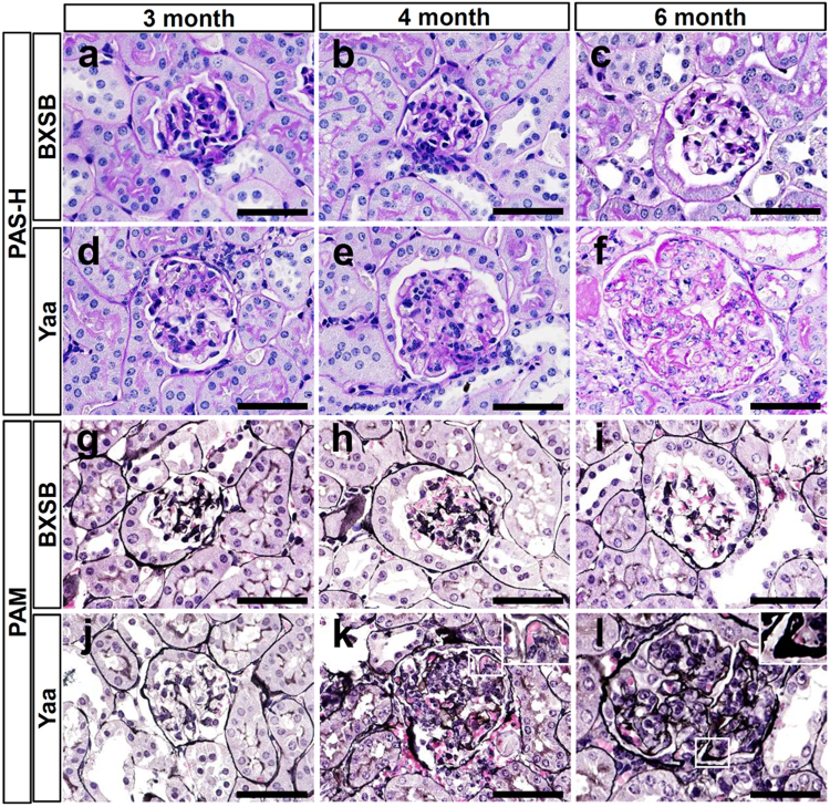
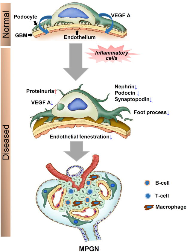
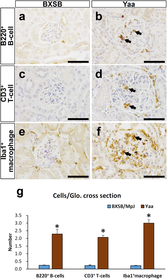
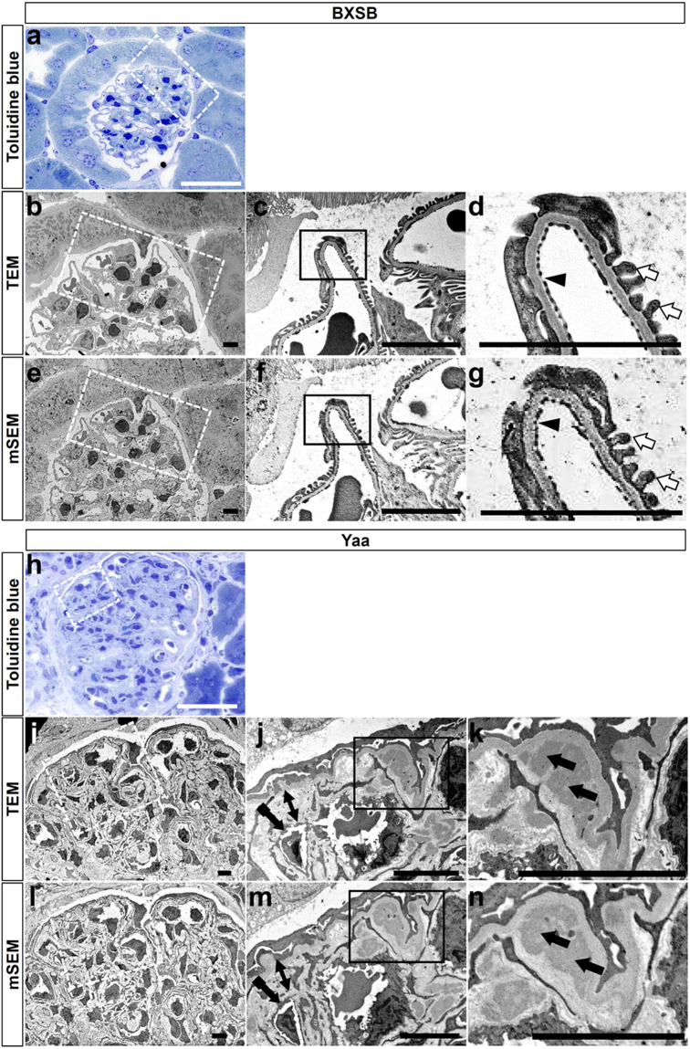
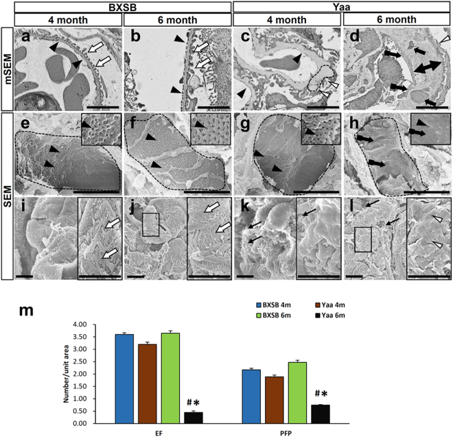
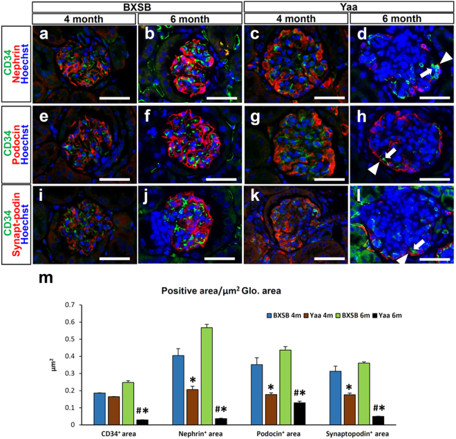
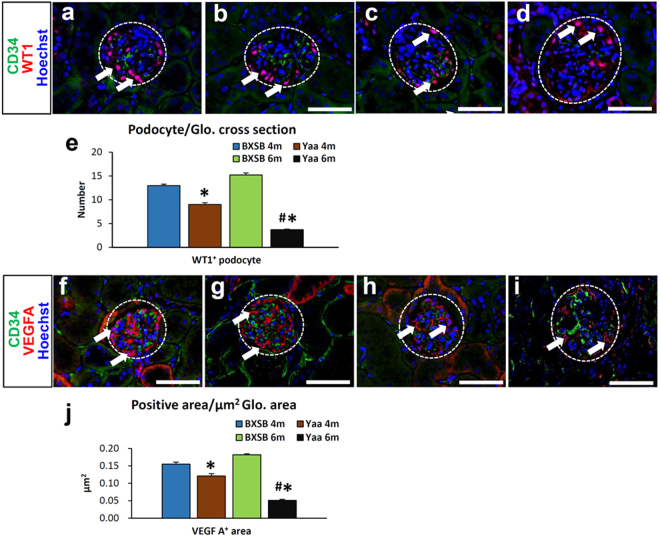
This study evaluated endothelial cells and podocytes, both being primary components of the glomerular filtration barrier, in the progression of membranoproliferative glomerulonephritis (MPGN) using modified scanning electron microscopy (mSEM) analysis. BXSB/MpJ-Yaa model mice exhibited autoimmune-mediated MPGN characterised by elevated serum autoantibody levels, albuminuria, renal dysfunctional parameters, and decreased glomerular endothelial fenestrations (EF) and podocyte foot process (PFP) effacement with immune cell infiltration. Similar to transmission electron microscopy, mSEM revealed a series of pathological changes in basement membrane and densities of EF and PFP in BXSB/MpJ-Yaa compared with control BXSB/MpJ at different stages. Further, immunopositive area of endothelial marker (CD34), podocyte functional molecules (Nephrin, Podocin, Synaptopodin, and Wilms' tumour 1 (WT1)), and vascular endothelial growth factor A (VEGF A) significantly decreased in the glomerulus of BXSB/MpJ-Yaa compared with BXSB at final stage. The indices of glomerular endothelial injuries (EF density and immunopositive area of CD34 and VEGF A) and podocyte injuries (PEP density and immunopositive area of podocyte functional molecules) were also significantly correlated with each other and with indices of autoimmune disease and renal dysfunction. Thus, our results elucidated the pathological crosstalk between endothelial cells and podocytes in MPGN progression and the usefulness of mSEM for glomerular pathological analysis.
本研究使用改良扫描电子显微镜(mSEM)分析评估了在膜增生性肾小球肾炎(MPGN)进展过程中内皮细胞和足细胞,这两种细胞都是肾小球滤过屏障的主要组成部分。BXSB/MpJ-Yaa 模型小鼠表现出自免疫介导的 MPGN,其特征是血清自身抗体水平升高、蛋白尿、肾功能参数降低以及肾小球内皮窗孔(EF)和足细胞足突融合(PFP)消失伴有免疫细胞浸润。与透射电子显微镜相似,mSEM 揭示了 BXSB/MpJ-Yaa 与对照 BXSB/MpJ 在不同阶段的基底膜以及 EF 和 PFP 密度的一系列病理变化。此外,与 BXSB 相比,在终末期 BXSB/MpJ-Yaa 肾小球中内皮标记物(CD34)、足细胞功能分子(Nephrin、Podocin、Synaptopodin 和 Wilms 肿瘤 1(WT1))和血管内皮生长因子 A(VEGF A)的免疫阳性面积显著降低。肾小球内皮损伤(EF 密度和 CD34 和 VEGF A 的免疫阳性面积)和足细胞损伤(PEP 密度和足细胞功能分子的免疫阳性面积)的指数也与自身免疫疾病和肾功能障碍的指数显著相关。因此,我们的结果阐明了 MPGN 进展中内皮细胞和足细胞之间的病理串扰以及 mSEM 对肾小球病理分析的有用性。