Xiao Yong-Bo, Cai Shao-Hang, Liu Li-Li, Yang Xia, Yun Jing-Ping
State Key Laboratory of Oncology in South China, Collaborative Innovation Center for Cancer Medicine, Sun Yat-sen University Cancer Center, Guangzhou 510060, China,
Department of Pathology, Sun Yat-sen University Cancer Center, Guangzhou 510060, China,
Cancer Manag Res. 2018 Jun 26;10:1781-1789. doi: 10.2147/CMAR.S166971. eCollection 2018.
Hepatocellular carcinoma (HCC) has a close relationship with lipid metabolism. Peroxisome proliferator-activated receptor α (PPARα) plays a crucial role in the regulation of fatty acid oxidation in the liver. However, the role of PPARα in HCC remains unclear.
A total of 804 HCC specimens were collected to construct a tissue microarray and for immunohistochemical analysis. The relationship between PPARα expression and clinical features of HCC patients was analyzed. Kaplan-Meier analysis was conducted to assess the prognostic value of PPARα expression levels.
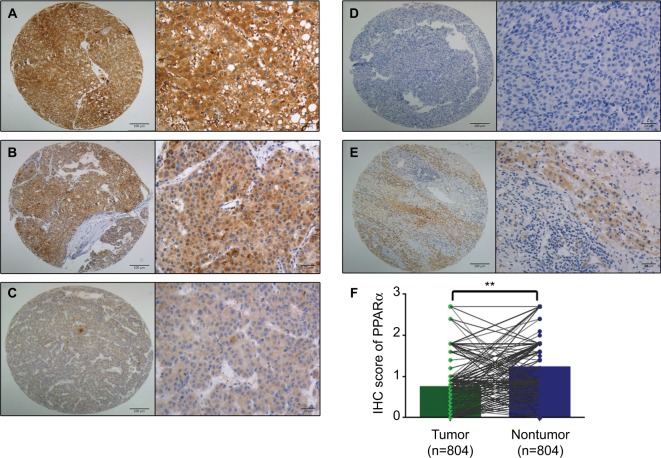
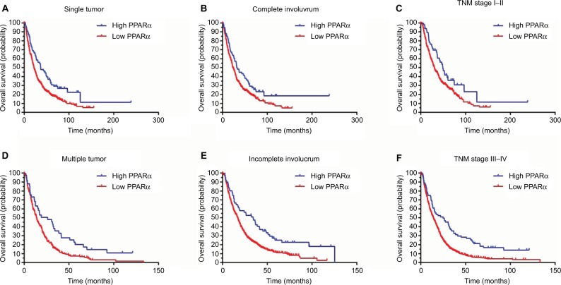
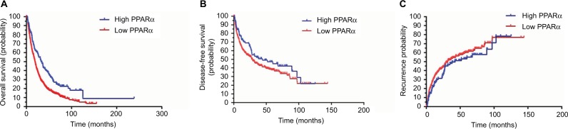
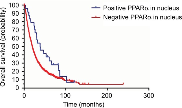
The expression of PPARα in HCC was noticeably decreased in HCC tissues. HCC patients with high levels of PPARα expression in cytoplasm had smaller tumors (=0.027), less vascular invasion (=0.049), and a higher proportion of complete involucrum (=0.038). Kaplan-Meier analysis showed that HCC patients with low PPARα expression in the cytoplasm had significantly worse outcomes in terms of overall survival (<0.001), disease-free survival (=0.024), and the probability of recurrence (=0.037). Similarly, overall survival was significantly shorter in HCC patients with negative PPARα expression in the nucleus (=0.034). Multivariate Cox analyses indicated that tumor size (=0.001), TNM stage (<0.001), vascular invasion (<0.001), and PPARα expression in the cytoplasm (<0.001) were found to be independent prognostic variables for overall survival.
Our data revealed that PPARα expression was decreased in HCC samples. High PPARα expression was correlated with longer survival times in HCC patients, and served as an independent factor for better outcomes. Our study therefore provides a promising biomarker for prognostic prediction and a potential therapeutic target for HCC.
肝细胞癌(HCC)与脂质代谢密切相关。过氧化物酶体增殖物激活受体α(PPARα)在肝脏脂肪酸氧化调节中起关键作用。然而,PPARα在HCC中的作用仍不清楚。
共收集804例HCC标本用于构建组织芯片及免疫组化分析。分析PPARα表达与HCC患者临床特征之间的关系。采用Kaplan-Meier分析评估PPARα表达水平的预后价值。
HCC组织中PPARα的表达明显降低。细胞质中PPARα表达水平高的HCC患者肿瘤较小(=0.027)、血管侵犯较少(=0.049)且完整包膜比例较高(=0.038)。Kaplan-Meier分析显示,细胞质中PPARα表达低的HCC患者在总生存期(<0.001)、无病生存期(=0.024)和复发概率(=0.037)方面的预后明显较差。同样,细胞核中PPARα表达阴性的HCC患者总生存期明显较短(=0.034)。多变量Cox分析表明,肿瘤大小(=0.001)、TNM分期(<0.001)、血管侵犯(<0.001)和细胞质中PPARα表达(<0.001)是总生存期的独立预后变量。
我们的数据显示HCC样本中PPARα表达降低。PPARα高表达与HCC患者较长的生存时间相关,是预后较好的独立因素。因此,我们的研究为预后预测提供了一个有前景的生物标志物,并为HCC提供了一个潜在的治疗靶点。