Department of Neurosurgery, Universitätsmedizin Charite - Campus Mitte, Luisenstrasse 46, 10117, Berlin, Germany.
Angiogenesis. 2018 Nov;21(4):873-881. doi: 10.1007/s10456-018-9633-6. Epub 2018 Jul 10.
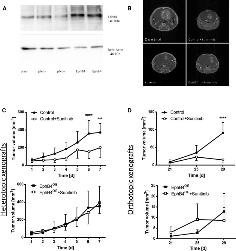
Alterations in vascular morphogenesis are hallmarks of antiangiogenesis-resistant tumor vessels. Vascular morphogenesis is regulated by ephrinB2-EphB4 system which may induce different biological effects depending on the oncological and molecular contexts. It was the aim of the current study to characterize the influence of EphB4 on tumor microcirculation after antiangiogenic treatment using different SF126 glioma models.
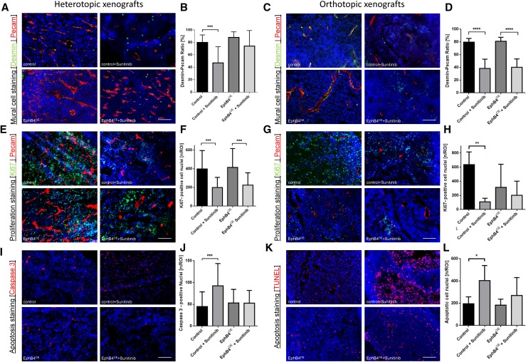
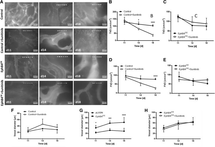
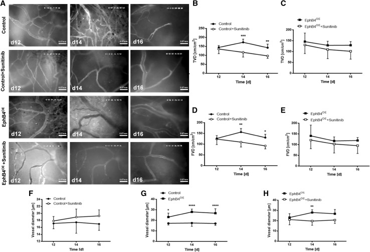
Using an ecotropic transfection system, empty vector (pLXSN) or EphB4 (EphB4) overexpressing Phoenix-ECO cells were coimplanted with SF126 glioma cells subcutaneously (dorsal skinfold chamber, DSC) and orthotopically (cranial window, CW). Tumor volume was assessed by MRI. Intravital microscopy (IVM) allowed microcirculatory analysis (total {TVD} and functional vessel density {FVD}, diameter {D}, and permeability index {PI}) before and after antiangiogenic treatment (Sunitinib: DSC: 40 mg/kg BW, 6 days; CW: 80 mg/kg BW, 4 days). Immunohistochemistry included Pecam-Desmin, Ki67, TUNEL, and Caspase 3 stainings.
EphB4 induced large and treatment-resistant tumor vessels (FVD: Control/Su: 110 ± 23 cm/cm vs. EphB4/Su: 103 ± 42 cm/cm). Maintenance of pericyte-endothelial cell interactions (Control: 80 ± 12 vs. Control/Su: 47 ± 26%; EphB4: 88 ± 9 vs. EphB4/Su: 74 ± 25%) and reduced antiproliferative (Control: 637 ± 80 vs. Control/Su: 110 ± 22; EphB4: 298 ± 108 vs. EphB4/Su: 213 ± 80) and proapoptotic responses (Control: 196 ± 25 vs. Control / Su: 404 ± 60; EphB4: 183 ± 20 vs. EphB4/Su: 270 ± 66) were observed under EphB4 overexpression.
EphB4 overexpression leads to vascular resistance by altering vascular morphogenesis, pericyte coverage, and cellular proliferation/apoptosis in experimental SF126 glioma models.
血管形态发生的改变是抗血管生成耐药肿瘤血管的标志。 EphrinB2-EphB4 系统调节血管形态发生,根据肿瘤学和分子背景,可能诱导不同的生物学效应。本研究旨在使用不同的 SF126 神经胶质瘤模型,描述 EphB4 对抗血管生成治疗后肿瘤微循环的影响。
使用外显子转染系统,空载体(pLXSN)或 EphB4(EphB4)过表达 Phoenix-ECO 细胞与 SF126 神经胶质瘤细胞皮下(背部皮肤囊,DSC)和原位(颅窗,CW)共植入。通过 MRI 评估肿瘤体积。在抗血管生成治疗(Sunitinib:DSC:40mg/kgBW,6 天;CW:80mg/kgBW,4 天)前后,通过活体显微镜(IVM)进行微血管分析(总 TVD 和功能血管密度 FVD、直径 D 和通透性指数 PI)。免疫组织化学包括 Pecam-Desmin、Ki67、TUNEL 和 Caspase 3 染色。
EphB4 诱导大的且对治疗有抗性的肿瘤血管(FVD:对照/Sunitinib:110±23cm/cm 与 EphB4/Sunitinib:103±42cm/cm)。维持周细胞-内皮细胞相互作用(对照:80±12 与对照/Sunitinib:47±26%;EphB4:88±9 与 EphB4/Sunitinib:74±25%)和减少增殖(对照:637±80 与对照/Sunitinib:110±22;EphB4:298±108 与 EphB4/Sunitinib:213±80)和促凋亡反应(对照:196±25 与对照/Sunitinib:404±60;EphB4:183±20 与 EphB4/Sunitinib:270±66)在 EphB4 过表达下观察到。
EphB4 过表达通过改变血管形态发生、周细胞覆盖和细胞增殖/凋亡,导致实验性 SF126 神经胶质瘤模型中的血管阻力。