• 文献检索
  • 文档翻译
  • 深度研究
  • 学术资讯
  • Suppr Zotero 插件Zotero 插件
  • 邀请有礼
  • 套餐&价格
  • 历史记录
应用&插件
Suppr Zotero 插件Zotero 插件浏览器插件Mac 客户端Windows 客户端微信小程序
定价
高级版会员购买积分包购买API积分包
服务
文献检索文档翻译深度研究API 文档MCP 服务
关于我们
关于 Suppr公司介绍联系我们用户协议隐私条款
关注我们

Suppr 超能文献

核心技术专利:CN118964589B侵权必究
粤ICP备2023148730 号-1Suppr @ 2026

文献检索

告别复杂PubMed语法,用中文像聊天一样搜索,搜遍4000万医学文献。AI智能推荐,让科研检索更轻松。

立即免费搜索

文件翻译

保留排版,准确专业,支持PDF/Word/PPT等文件格式,支持 12+语言互译。

免费翻译文档

深度研究

AI帮你快速写综述,25分钟生成高质量综述,智能提取关键信息,辅助科研写作。

立即免费体验

蛛网膜和软脑膜细胞中的 VE-cadherin 可作为 CNS 免疫监视和炎症的体内成像的合适标志物。

VE-cadherin in arachnoid and pia mater cells serves as a suitable landmark for in vivo imaging of CNS immune surveillance and inflammation.

机构信息

Theodor Kocher Institute, University of Bern, Bern, Switzerland.

Department of Immunology, Genetics and Pathology, Uppsala University, Uppsala, Sweden.

出版信息

Nat Commun. 2023 Sep 20;14(1):5837. doi: 10.1038/s41467-023-41580-4.

DOI:10.1038/s41467-023-41580-4
PMID:37730744
原文链接:https://pmc.ncbi.nlm.nih.gov/articles/PMC10511632/
Abstract

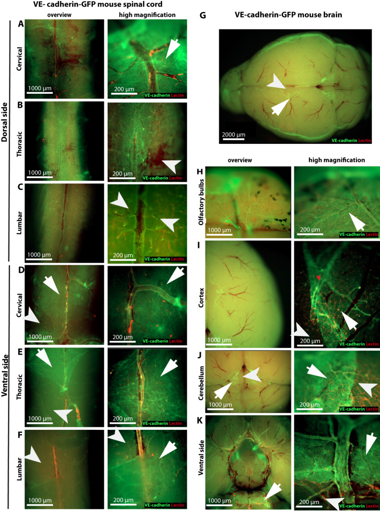
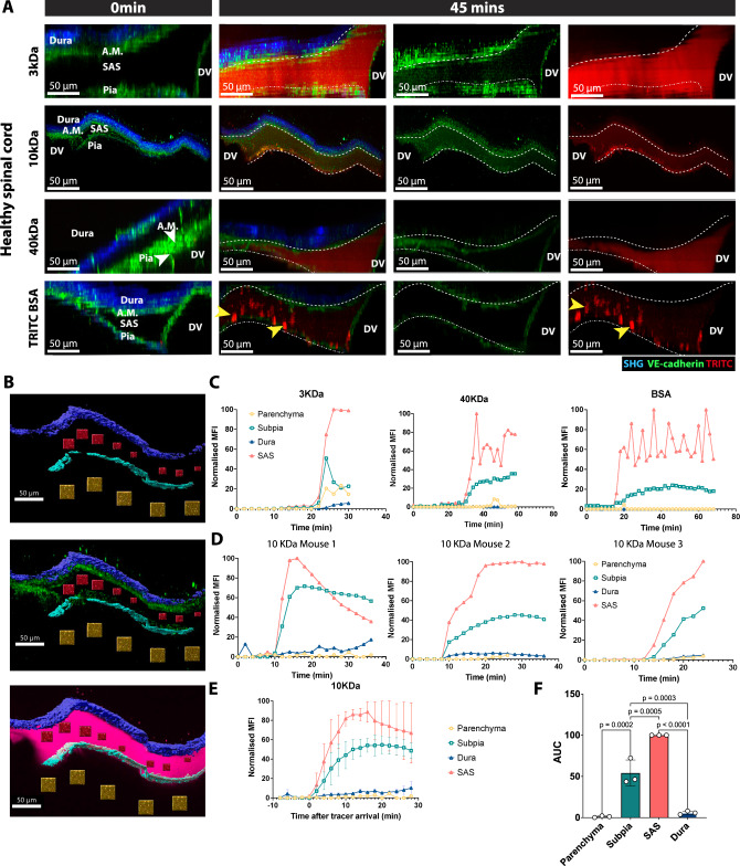
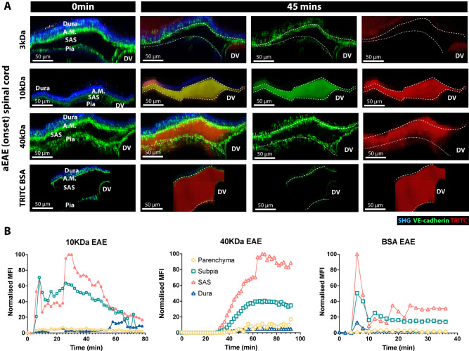
Meninges cover the surface of the brain and spinal cord and contribute to protection and immune surveillance of the central nervous system (CNS). How the meningeal layers establish CNS compartments with different accessibility to immune cells and immune mediators is, however, not well understood. Here, using 2-photon imaging in female transgenic reporter mice, we describe VE-cadherin at intercellular junctions of arachnoid and pia mater cells that form the leptomeninges and border the subarachnoid space (SAS) filled with cerebrospinal fluid (CSF). VE-cadherin expression also marked a layer of Prox1 cells located within the arachnoid beneath and separate from E-cadherin arachnoid barrier cells. In vivo imaging of the spinal cord and brain in female VE-cadherin-GFP reporter mice allowed for direct observation of accessibility of CSF derived tracers and T cells into the SAS bordered by the arachnoid and pia mater during health and neuroinflammation, and detection of volume changes of the SAS during CNS pathology. Together, the findings identified VE-cadherin as an informative landmark for in vivo imaging of the leptomeninges that can be used to visualize the borders of the SAS and thus potential barrier properties of the leptomeninges in controlling access of immune mediators and immune cells into the CNS during health and neuroinflammation.

摘要

脑膜覆盖在大脑和脊髓表面,有助于保护和监测中枢神经系统(CNS)的免疫。然而,脑膜各层如何建立起具有不同免疫细胞和免疫介质可及性的 CNS 隔室尚不清楚。在这里,我们使用雌性转基因报告小鼠的双光子成像,描述了位于软脑膜和软膜细胞(形成软脑膜)的细胞间连接处的 VE-钙黏蛋白,以及充满脑脊液(CSF)的蛛网膜下腔(SAS)的边界。VE-钙黏蛋白的表达也标记了位于蛛网膜下腔下方的 Prox1 细胞层,与位于蛛网膜下腔的 E-钙黏蛋白蛛网膜屏障细胞分开。在雌性 VE-钙黏蛋白-GFP 报告小鼠的脊髓和大脑的体内成像中,我们可以直接观察到 CSF 衍生示踪剂和 T 细胞在健康和神经炎症期间进入由软膜和软膜边界的 SAS,以及在 CNS 病理期间检测到 SAS 体积的变化。总之,这些发现确定了 VE-钙黏蛋白是用于体内成像软脑膜的信息性标志物,可用于可视化 SAS 的边界,从而控制在健康和神经炎症期间免疫介质和免疫细胞进入 CNS 的能力。

https://cdn.ncbi.nlm.nih.gov/pmc/blobs/80b3/10511632/3e02ac17a866/41467_2023_41580_Fig10_HTML.jpg
https://cdn.ncbi.nlm.nih.gov/pmc/blobs/80b3/10511632/b11094ca1f7e/41467_2023_41580_Fig1_HTML.jpg
https://cdn.ncbi.nlm.nih.gov/pmc/blobs/80b3/10511632/7f49d4a47c98/41467_2023_41580_Fig2_HTML.jpg
https://cdn.ncbi.nlm.nih.gov/pmc/blobs/80b3/10511632/09dacec4f806/41467_2023_41580_Fig3_HTML.jpg
https://cdn.ncbi.nlm.nih.gov/pmc/blobs/80b3/10511632/845240dacd60/41467_2023_41580_Fig4_HTML.jpg
https://cdn.ncbi.nlm.nih.gov/pmc/blobs/80b3/10511632/8ab40c54c306/41467_2023_41580_Fig5_HTML.jpg
https://cdn.ncbi.nlm.nih.gov/pmc/blobs/80b3/10511632/1ab10231ce06/41467_2023_41580_Fig6_HTML.jpg
https://cdn.ncbi.nlm.nih.gov/pmc/blobs/80b3/10511632/ba6dc37a67cd/41467_2023_41580_Fig7_HTML.jpg
https://cdn.ncbi.nlm.nih.gov/pmc/blobs/80b3/10511632/4a8b54c133bc/41467_2023_41580_Fig8_HTML.jpg
https://cdn.ncbi.nlm.nih.gov/pmc/blobs/80b3/10511632/5b59516fc48a/41467_2023_41580_Fig9_HTML.jpg
https://cdn.ncbi.nlm.nih.gov/pmc/blobs/80b3/10511632/3e02ac17a866/41467_2023_41580_Fig10_HTML.jpg

相似文献

1
VE-cadherin in arachnoid and pia mater cells serves as a suitable landmark for in vivo imaging of CNS immune surveillance and inflammation.蛛网膜和软脑膜细胞中的 VE-cadherin 可作为 CNS 免疫监视和炎症的体内成像的合适标志物。
Nat Commun. 2023 Sep 20;14(1):5837. doi: 10.1038/s41467-023-41580-4.
2
Arachnoiditis蛛网膜炎
3
Postnatal meningeal CSF transport is primarily mediated by the arachnoid and pia maters and is not altered after intraventricular hemorrhage-posthemorrhagic hydrocephalus.产后脑膜 CSF 转运主要由软脑膜和脑被膜介导,且在脑室出血-出血后脑积水后不会改变。
Fluids Barriers CNS. 2024 Jan 8;21(1):4. doi: 10.1186/s12987-023-00503-7.
4
Molecular anatomy of adult mouse leptomeninges.成年小鼠软脑膜的分子解剖结构。
Neuron. 2023 Dec 6;111(23):3745-3764.e7. doi: 10.1016/j.neuron.2023.09.002. Epub 2023 Sep 29.
5
The cranial arachnoid and pia mater in man: anatomical and ultrastructural observations.人类颅蛛网膜和软脑膜:解剖学与超微结构观察
Neuropathol Appl Neurobiol. 1988 Jan-Feb;14(1):1-17. doi: 10.1111/j.1365-2990.1988.tb00862.x.
6
Microscopic morphology and histology of the human meninges.人类脑膜的微观形态学与组织学
Morphologie. 2005 Mar;89(284):22-34. doi: 10.1016/s1286-0115(05)83235-7.
7
Anatomy, Head and Neck, Subarachnoid Space解剖学,头颈部,蛛网膜下腔
8
Commentary on "Structural characterization of SLYM - a 4th meningeal membrane".对“SLYM——第四脑膜的结构特征”的评论。
Fluids Barriers CNS. 2024 Sep 9;21(1):69. doi: 10.1186/s12987-024-00568-y.
9
Viscoelasticity of spinal cord and meningeal tissues.脊髓和脑膜组织的粘弹性。
Acta Biomater. 2018 Jul 15;75:253-262. doi: 10.1016/j.actbio.2018.05.045. Epub 2018 May 28.
10
Morphine and alfentanil permeability through the spinal dura, arachnoid, and pia mater of dogs and monkeys.吗啡和阿芬太尼透过犬和猴的脊髓硬脑膜、蛛网膜和软脑膜的通透性。
Anesthesiology. 1990 Dec;73(6):1214-9. doi: 10.1097/00000542-199012000-00020.

引用本文的文献

1
A comprehensive molecular atlas of the mesenchymal cell types in the mouse liver.小鼠肝脏间充质细胞类型的综合分子图谱。
EMBO Rep. 2025 Sep 15. doi: 10.1038/s44319-025-00580-9.
2
Skull-meninges-brain connectivity and extra-axial brain tumours.颅骨-脑膜-脑连接与脑外肿瘤
Brain Commun. 2025 Aug 21;7(5):fcaf311. doi: 10.1093/braincomms/fcaf311. eCollection 2025.
3
Cerebrospinal fluid draining lymphatics in health and disease: advances and controversies.健康与疾病状态下的脑脊液引流淋巴管:进展与争议

本文引用的文献

1
Antigen recognition detains CD8 T cells at the blood-brain barrier and contributes to its breakdown.抗原识别将 CD8 T 细胞滞留在血脑屏障处,并导致其破坏。
Nat Commun. 2023 May 30;14(1):3106. doi: 10.1038/s41467-023-38703-2.
2
Formation and function of the meningeal arachnoid barrier around the developing mouse brain.发育中小鼠脑周围脑膜蛛网膜屏障的形成和功能。
Dev Cell. 2023 Apr 24;58(8):635-644.e4. doi: 10.1016/j.devcel.2023.03.005. Epub 2023 Mar 29.
3
A mesothelium divides the subarachnoid space into functional compartments.
Nat Cardiovasc Res. 2025 Sep 8. doi: 10.1038/s44161-025-00705-2.
4
Identifying a potential role of immune cells in gadolinium deposition within the brain.确定免疫细胞在大脑钆沉积中的潜在作用。
Fluids Barriers CNS. 2025 Jul 29;22(1):80. doi: 10.1186/s12987-025-00674-5.
5
Role of CSF flow and meningeal barriers in the development of inflammatory lesions at the CNS-PNS transition zone of cranial nerves in autoimmune demyelinating diseases.脑脊液流动和脑膜屏障在自身免疫性脱髓鞘疾病中脑神经中枢神经系统-外周神经系统过渡区炎性病变发展中的作用。
Acta Neuropathol. 2025 Jun 19;149(1):65. doi: 10.1007/s00401-025-02896-1.
6
Brain fluid physiology in ischaemic stroke; more than just oedema.缺血性卒中中的脑脊液生理学;不止于水肿。
Fluids Barriers CNS. 2025 Jun 18;22(1):60. doi: 10.1186/s12987-025-00671-8.
7
A route for cerebrospinal fluid flow through leptomeningeal arterial-venous overlaps enables macromolecule and fluid shunting.脑脊液通过软脑膜动静脉重叠处的流动途径可实现大分子和液体分流。
Nat Neurosci. 2025 Jun 9. doi: 10.1038/s41593-025-01977-4.
8
Lack of junctional adhesion molecule (JAM)-B traps CD8 T cells in CNS border zones and ameliorates autoimmune neuroinflammation.缺乏连接粘附分子(JAM)-B会将CD8 T细胞困在中枢神经系统边界区域,并改善自身免疫性神经炎症。
Acta Neuropathol Commun. 2025 May 26;13(1):117. doi: 10.1186/s40478-025-02021-z.
9
CSF transport at the brain-meningeal border: effects on neurological health and disease.脑脊液在脑-脑膜边界处的转运:对神经健康和疾病的影响。
Lancet Neurol. 2025 Jun;24(6):535-547. doi: 10.1016/S1474-4422(25)00115-2.
10
Border-associated macrophages as gatekeepers of brain homeostasis and immunity.边界相关巨噬细胞作为脑稳态和免疫的守门人。
Immunity. 2025 May 13;58(5):1085-1100. doi: 10.1016/j.immuni.2025.04.005. Epub 2025 May 4.
间皮将蛛网膜下腔分隔成不同的功能腔室。
Science. 2023 Jan 6;379(6627):84-88. doi: 10.1126/science.adc8810. Epub 2023 Jan 5.
4
Cerebrospinal fluid regulates skull bone marrow niches via direct access through dural channels.脑脊液通过硬脑膜通道直接进入调节颅骨骨髓龛。
Nat Neurosci. 2022 May;25(5):555-560. doi: 10.1038/s41593-022-01029-1. Epub 2022 Mar 17.
5
How Does the Immune System Enter the Brain?免疫系统如何进入大脑?
Front Immunol. 2022 Feb 22;13:805657. doi: 10.3389/fimmu.2022.805657. eCollection 2022.
6
A human blood-arachnoid barrier atlas of transporters, receptors, enzymes, and tight junction and marker proteins: Comparison with dog and pig in absolute abundance.人类血脑屏障转运体、受体、酶、紧密连接和标记蛋白图谱:与犬和猪的绝对丰度比较。
J Neurochem. 2022 Apr;161(2):187-208. doi: 10.1111/jnc.15599. Epub 2022 Mar 17.
7
Central nervous system zoning: How brain barriers establish subdivisions for CNS immune privilege and immune surveillance.中枢神经系统分区:脑屏障如何为中枢神经系统的免疫特权和免疫监视建立细分。
J Intern Med. 2022 Jul;292(1):47-67. doi: 10.1111/joim.13469. Epub 2022 Mar 14.
8
Emerging roles for CNS fibroblasts in health, injury and disease.中枢神经系统成纤维细胞在健康、损伤和疾病中的新作用。
Nat Rev Neurosci. 2022 Jan;23(1):23-34. doi: 10.1038/s41583-021-00525-w. Epub 2021 Oct 20.
9
Single-cell profiling of CNS border compartment leukocytes reveals that B cells and their progenitors reside in non-diseased meninges.对中枢神经系统边界隔室白细胞进行单细胞分析显示,B 细胞及其前体细胞存在于非病变脑膜中。
Nat Neurosci. 2021 Sep;24(9):1225-1234. doi: 10.1038/s41593-021-00880-y. Epub 2021 Jul 12.
10
Heterogeneity of meningeal B cells reveals a lymphopoietic niche at the CNS borders.脑膜 B 细胞的异质性揭示了 CNS 边界处的淋系造血龛位。
Science. 2021 Jul 23;373(6553). doi: 10.1126/science.abf9277. Epub 2021 Jun 3.