Department of Neurosurgery, Medical University of Innsbruck, Anichstrasse 35, 6020, Innsbruck, Austria.
Department of Neurosurgery, Klinikum rechts der Isar, Technische Universität München, Munich, Germany.
J Neurooncol. 2018 Sep;139(3):699-711. doi: 10.1007/s11060-018-2916-3. Epub 2018 Jul 10.
Imaging studies in diffuse low-grade gliomas (DLGG) vary across centers. In order to establish a minimal core of imaging necessary for further investigations and clinical trials in the field of DLGG, we aimed to establish the status quo within specialized European centers.
An online survey composed of 46 items was sent out to members of the European Low-Grade Glioma Network, the European Association of Neurosurgical Societies, the German Society of Neurosurgery and the Austrian Society of Neurosurgery.
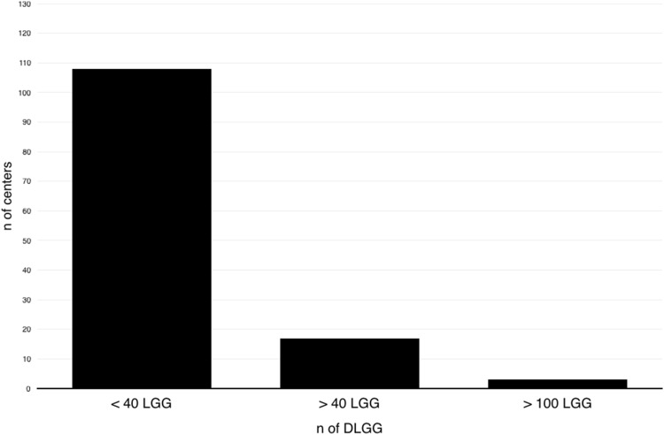
A total of 128 fully completed surveys were received and analyzed. Most centers (n = 96, 75%) were academic and half of the centers (n = 64, 50%) adhered to a dedicated treatment program for DLGG. There were national differences regarding the sequences enclosed in MRI imaging and use of PET, however most included T1 (without and with contrast, 100%), T2 (100%) and TIRM or FLAIR (20, 98%). DWI is performed by 80% of centers and 61% of centers regularly performed PWI.
A minimal core of imaging composed of T1 (w/wo contrast), T2, TIRM/FLAIR, PWI and DWI could be identified. All morphologic images should be obtained in a slice thickness of ≤ 3 mm. No common standard could be obtained regarding advanced MRI protocols and PET.
We believe that our study makes a significant contribution to the literature because we were able to determine similarities in numerous aspects of LGG imaging. Using the proposed "minimal core of imaging" in clinical routine will facilitate future cooperative studies.
弥漫性低级别胶质瘤(DLGG)的影像学研究在各中心之间存在差异。为了确定在 DLGG 领域进一步研究和临床试验所需的影像学最小核心,我们旨在确定专业欧洲中心的现状。
向欧洲低级别神经胶质瘤网络、欧洲神经外科学会、德国神经外科学会和奥地利神经外科学会的成员发送了一份包含 46 个项目的在线调查。
共收到并分析了 128 份完整填写的调查。大多数中心(n=96,75%)为学术性的,一半的中心(n=64,50%)遵循专门的 DLGG 治疗方案。在 MRI 成像中包含的序列和使用 PET 方面存在国家差异,然而大多数中心包括 T1(无对比和有对比,100%)、T2(100%)和 TIRM 或 FLAIR(20%,98%)。80%的中心进行 DWI,61%的中心定期进行 PWI。
可以确定由 T1(w/wo 对比)、T2、TIRM/FLAIR、PWI 和 DWI 组成的最小核心影像学。所有形态学图像的切片厚度应≤3mm。关于高级 MRI 协议和 PET 没有获得共同的标准。
我们认为我们的研究对文献做出了重要贡献,因为我们能够确定 LGG 影像学的许多方面存在相似之处。在临床常规中使用建议的“最小核心影像学”将促进未来的合作研究。