IEEE Trans Biomed Eng. 2019 Mar;66(3):647-655. doi: 10.1109/TBME.2018.2853571. Epub 2018 Jul 5.
To both qualitatively and quantitatively investigate corneal biomechanical properties through an ultrasonic microelastography imaging system, which is potentially useful in the diagnosis of diseases, such as keratoconus, postrefractive keratectasia, and tracking treatment such as cross-linking surgery.
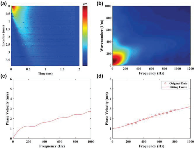
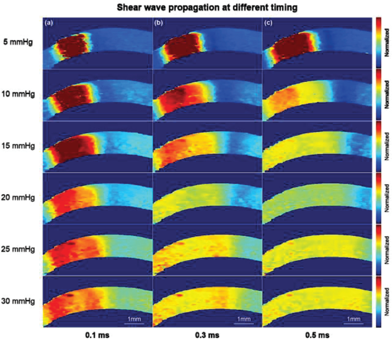
Our imaging system has a dual-frequency configuration, including a 4.5 MHz ring transducer to push the tissue and a confocally aligned 40 MHz needle transducer to track micron-level displacement. Two-dimensional/three-dimensional acoustic radiation force impulse (ARFI) imaging and Young's modulus in the region of interest were performed on ex vivo porcine corneas that were either cross-linked using formalin solution or preloaded with intraocular pressure (IOPs) from 5 to 30 mmHg.
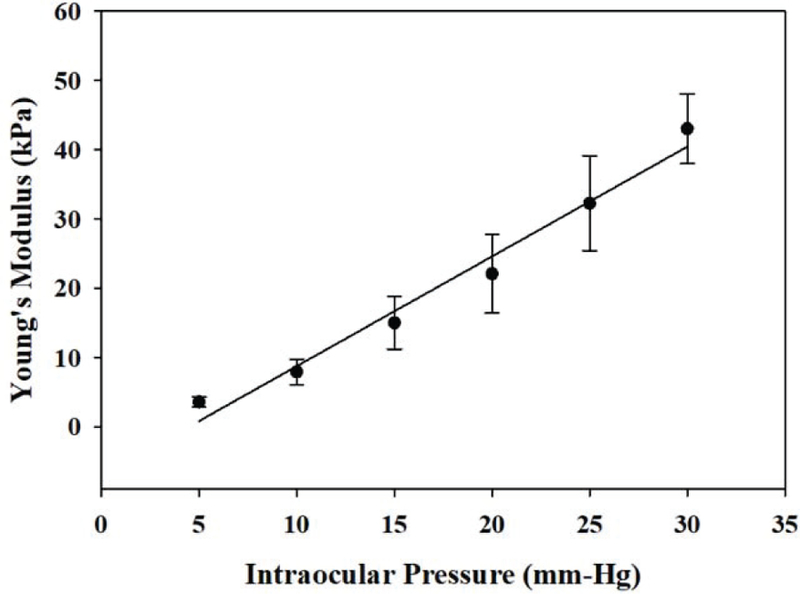
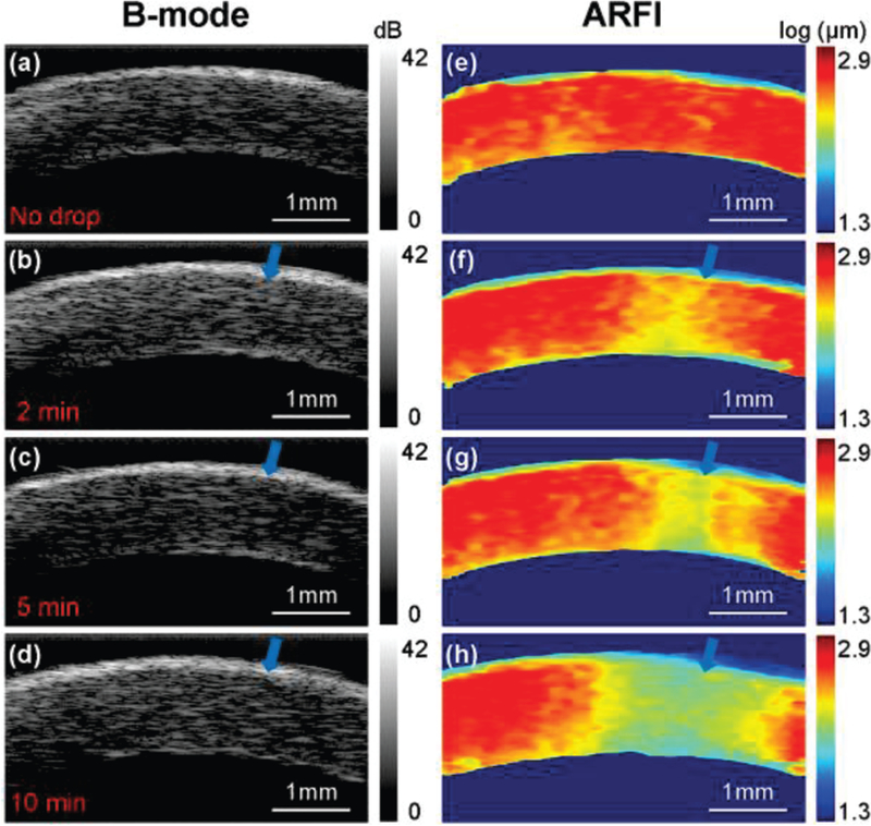
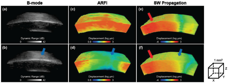
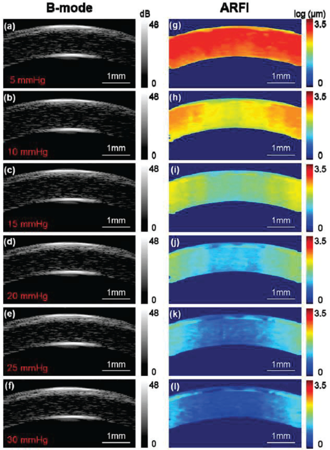
The increase of corneal stiffness and the change in cross-linked volume following formalin crosslinking could be precisely observed in the ARFI images and reflected by the reconstructed Young's modulus while the B-mode structural images remained almost unchanged. In addition, the relationship between the stiffness of the cornea and IOPs was investigated among 12 porcine corneas. The corneal stiffness is significantly different at various IOPs and has a tendency to become stiffer with increasing IOP.
Our results demonstrate the principle of using ultrasonic microelastography techniques to image the biomechanical properties of the cornea. Integrating high-resolution ARFI imaging labeled with reconstructed Young's modulus and structural imaging of the cornea can potentially lead to a routinely performed imaging modality in the field of ophthalmology.
通过超声微弹性成像系统对角膜生物力学特性进行定性和定量研究,该系统在诊断圆锥角膜、屈光性角膜后表面扩张等疾病以及交联手术等治疗效果的跟踪方面具有潜在的应用价值。
我们的成像系统采用双频配置,包括一个 4.5MHz 的环式换能器来推动组织和一个共焦对准的 40MHz 针式换能器来跟踪微米级的位移。对使用福尔马林溶液交联的离体猪眼角膜或从 5mmHg 到 30mmHg 加载眼内压(IOP)的离体猪眼角膜进行二维/三维声辐射力脉冲(ARFI)成像和感兴趣区域的杨氏模量测量。
福尔马林交联后角膜硬度的增加和交联体积的变化可以在 ARFI 图像中精确观察到,并反映在重建的杨氏模量中,而 B 模式结构图像几乎保持不变。此外,还研究了 12 只猪眼角膜之间的角膜硬度与 IOP 之间的关系。在不同的 IOP 下,角膜硬度有显著差异,并且随着 IOP 的增加,角膜硬度有变硬的趋势。
我们的结果表明,超声微弹性成像技术可用于成像角膜的生物力学特性。整合具有重建杨氏模量标记的高分辨率 ARFI 成像和角膜的结构成像,可能会成为眼科领域常规的成像方式。