USC Roski Eye Institute, University of Southern California, Los Angeles, CA, USA.
Department of Biomedical Engineering, University of Southern California, Los Angeles, CA, USA.
Sci Rep. 2017 Apr 27;7(1):1230. doi: 10.1038/s41598-017-01210-8.
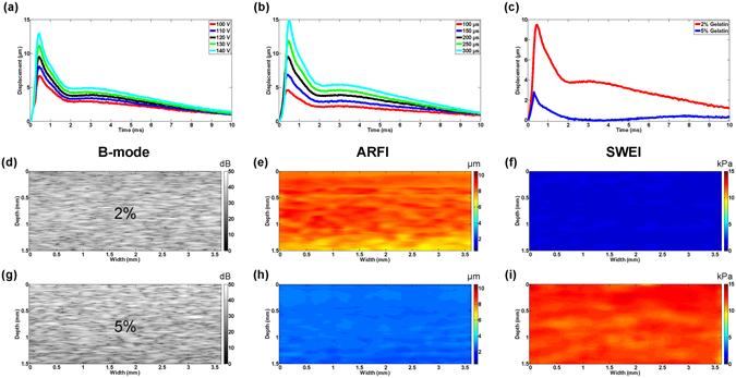
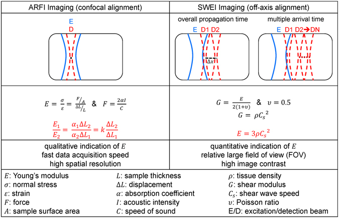
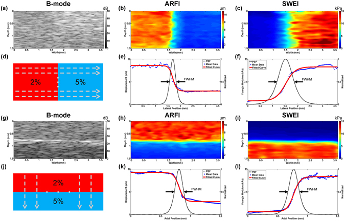
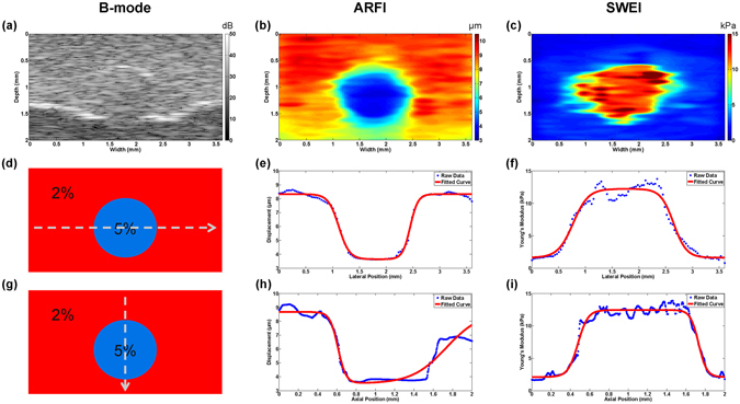
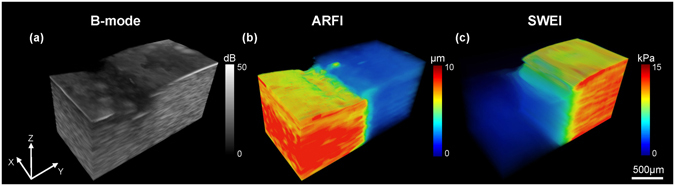
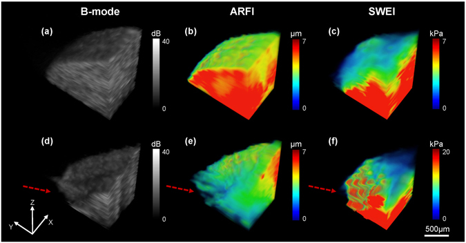
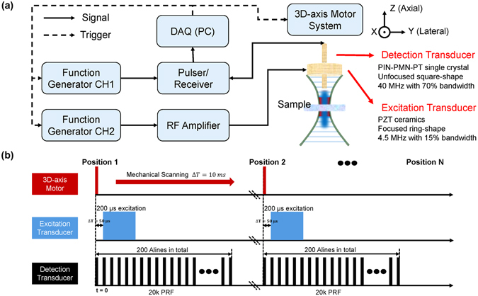
In clinical decision making, in addition to anatomical information, biomechanical properties of soft tissues may provide additional clues for disease diagnosis. Given the fact that most of diseases are originated from micron sized structures, an elastography imaging system of fine resolution (~100 µm) and deep penetration depth capable of providing both qualitative and quantitative measurements of biomechanical properties is desired. Here, we report a newly developed multi-functional ultrasonic micro-elastography imaging system in which acoustic radiation force impulse imaging (ARFI) and shear wave elasticity imaging (SWEI) are implemented. To accomplish this, the 4.5 MHz/40 MHz transducer were used as the excitation/detection source, respectively. The imaging system was tested with tissue-mimicking phantoms and an ex vivo chicken liver through 2D/3D imaging. The measured lateral/axial elastography resolution and field of view are 223.7 ± 20.1/109.8 ± 6.9 µm and 1.5 mm for ARFI, 543.6 ± 39.3/117.6 ± 8.7 µm and 2 mm for SWEI, respectively. These results demonstrate that the promising capability of this high resolution elastography imaging system for characterizing tissue biomechanical properties at microscale level and its translational potential into clinical practice.
在临床决策中,除了解剖信息外,软组织的生物力学特性也可为疾病诊断提供额外线索。鉴于大多数疾病都起源于微米级结构,因此需要一种具有精细分辨率(约 100μm)和深穿透深度的弹性成像系统,能够提供生物力学特性的定性和定量测量。在这里,我们报告了一种新开发的多功能超声微弹性成像系统,该系统实现了声辐射力脉冲成像(ARFI)和剪切波弹性成像(SWEI)。为此,分别使用 4.5MHz/40MHz 换能器作为激励/检测源。通过 2D/3D 成像,对组织模拟体模和离体鸡肝进行了成像系统测试。ARFI 的测量横向/轴向弹性成像分辨率和视野分别为 223.7±20.1/109.8±6.9μm 和 1.5mm,SWEI 的为 543.6±39.3/117.6±8.7μm 和 2mm。这些结果表明,该高分辨率弹性成像系统具有在微尺度上对组织生物力学特性进行特征描述的潜力,并且有可能转化为临床实践。