IEEE Trans Vis Comput Graph. 2019 Apr;25(4):1760-1773. doi: 10.1109/TVCG.2018.2818701. Epub 2018 Mar 27.
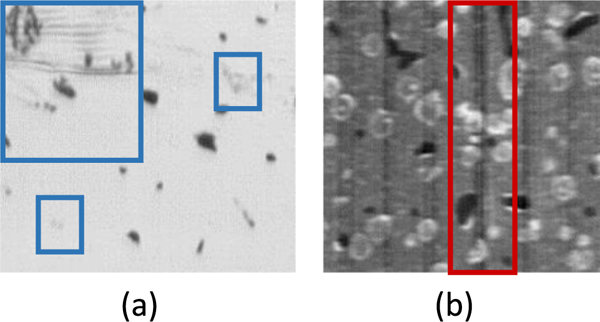
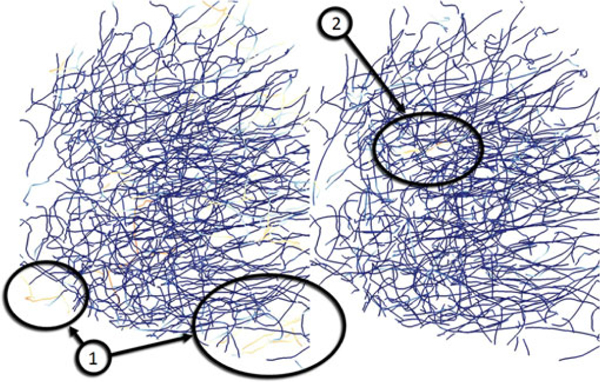
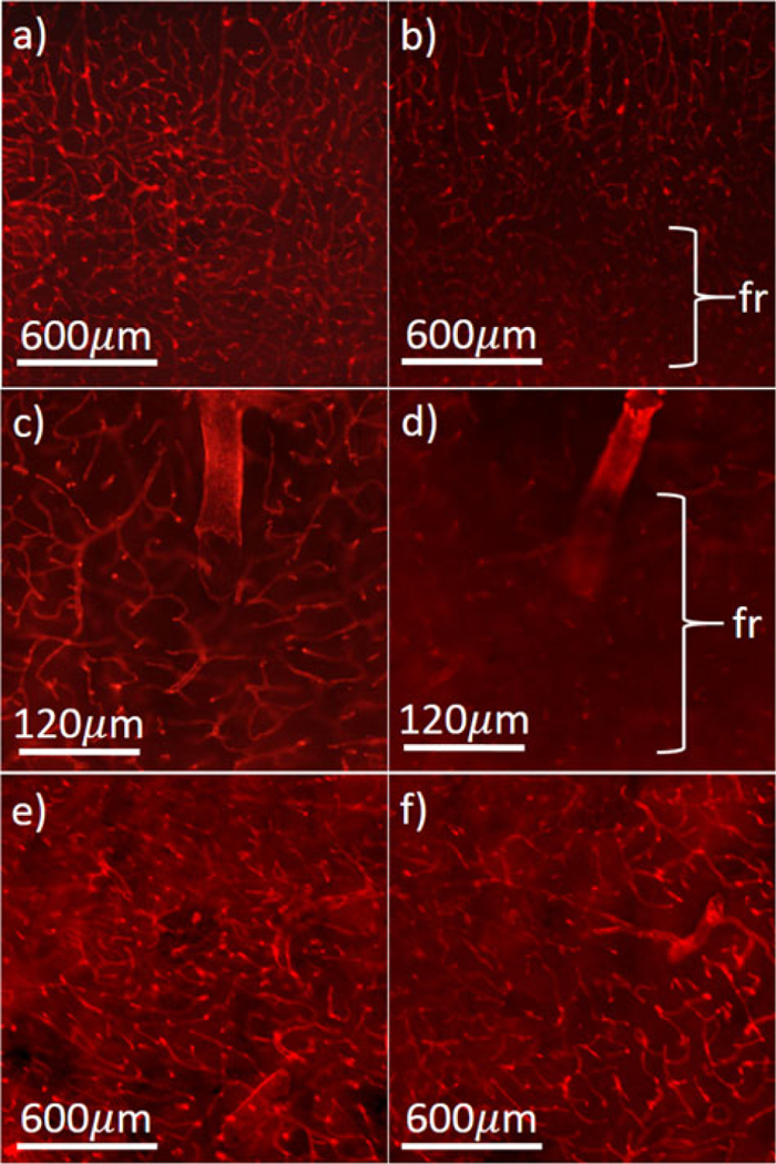
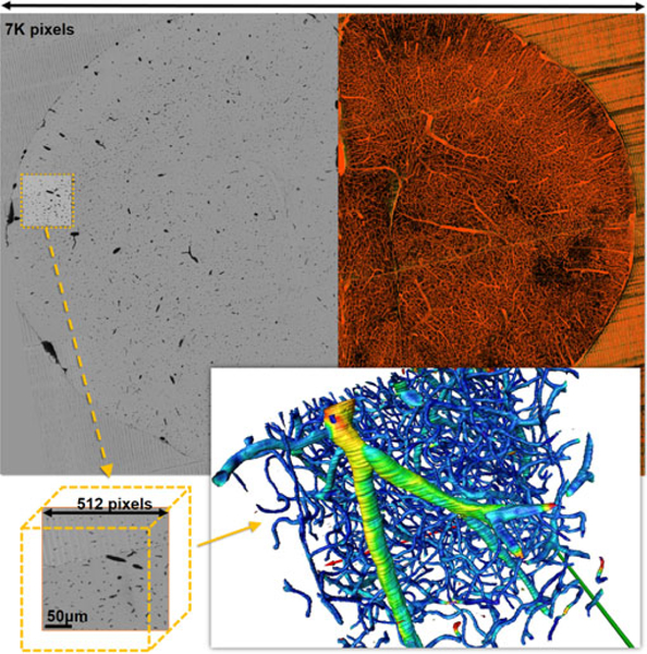
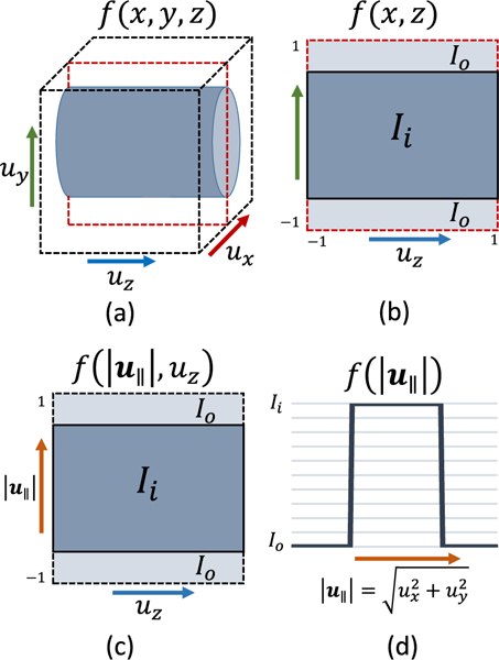
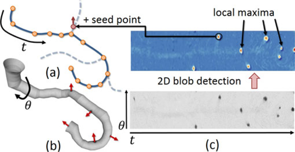
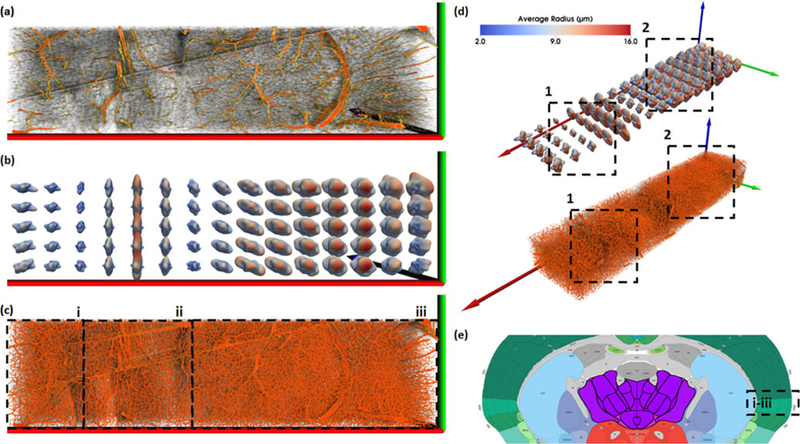
Advances in high-throughput imaging allow researchers to collect three-dimensional images of whole organ microvascular networks. These extremely large images contain networks that are highly complex, time consuming to segment, and difficult to visualize. In this paper, we present a framework for segmenting and visualizing vascular networks from terabyte-sized three-dimensional images collected using high-throughput microscopy. While these images require terabytes of storage, the volume devoted to the fiber network is ≈ 4 percent of the total volume size. While the networks themselves are sparse, they are tremendously complex, interconnected, and vary widely in diameter. We describe a parallel GPU-based predictor-corrector method for tracing filaments that is robust to noise and sampling errors common in these data sets. We also propose a number of visualization techniques designed to convey the complex statistical descriptions of fibers across large tissue sections-including commonly studied microvascular characteristics, such as orientation and volume.
高通量成像技术的进步使得研究人员能够采集整个器官微血管网络的三维图像。这些极其庞大的图像包含高度复杂的网络,需要花费大量时间进行分割,并且难以可视化。在本文中,我们提出了一种从使用高通量显微镜采集的兆字节大小的三维图像中分割和可视化血管网络的框架。虽然这些图像需要兆字节的存储空间,但纤维网络的体积仅占总体积大小的 ≈ 4%。虽然网络本身是稀疏的,但它们非常复杂,相互连接,并且直径差异很大。我们描述了一种基于 GPU 的并行预测校正方法,用于跟踪纤维,该方法对这些数据集常见的噪声和采样误差具有鲁棒性。我们还提出了许多可视化技术,旨在传达跨越大组织切片的纤维的复杂统计描述-包括通常研究的微血管特征,例如方向和体积。