Luna Arthur Cássio de Lima, Saraiva Greice Kelle Viegas, Chierice Gilberto Orivaldo, Hesse Henrique, Maria Durvanei Augusto
Department of Biochemistry and Biophysics, Butantan Institute, 1500, Vital Brasil Avenue, Sao Paulo, 05503-900, Brazil.
Department of Medical Sciences, Medical School, University of Sao Paulo, Sao Paulo, Brazil.
BMC Pharmacol Toxicol. 2018 Jul 11;19(1):44. doi: 10.1186/s40360-018-0225-2.
Current studies have demonstrated that DODAC/PHO-S (Dioctadecyldimethylammonium Chloride/Synthetic phosphoethanolamine) liposomes induces cytotoxicity in Hepa1c1c7 and B16F10 murine tumor cells, with a higher proportion than PHO-S. Therefore, our aim was to evaluate the potential of DODAC/PHO-S to elucidate the mechanism of cell death whereby the liposomes induces cytotoxicity in hepatocellular carcinoma Hepa1c1c7, compared to the PHO-S alone.
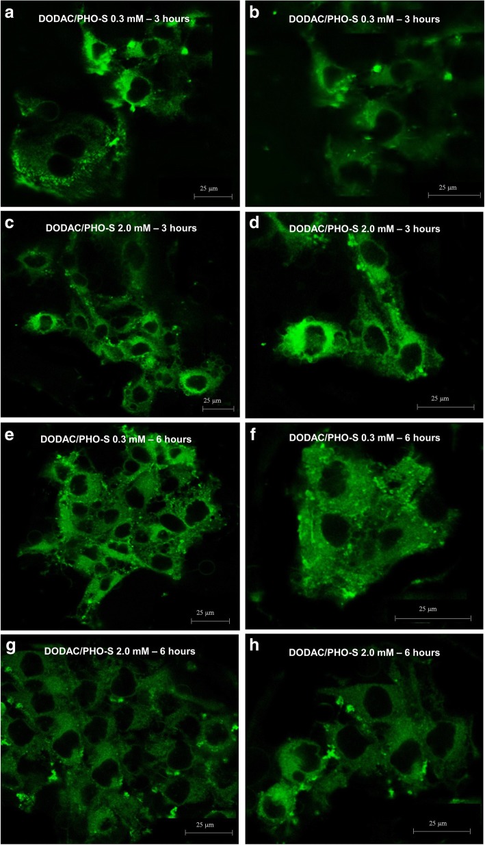
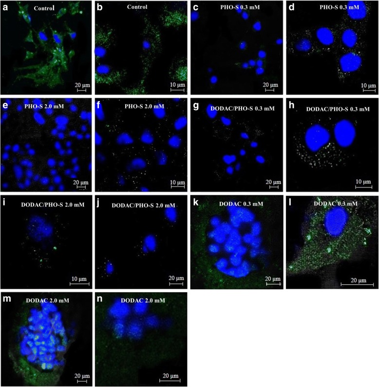
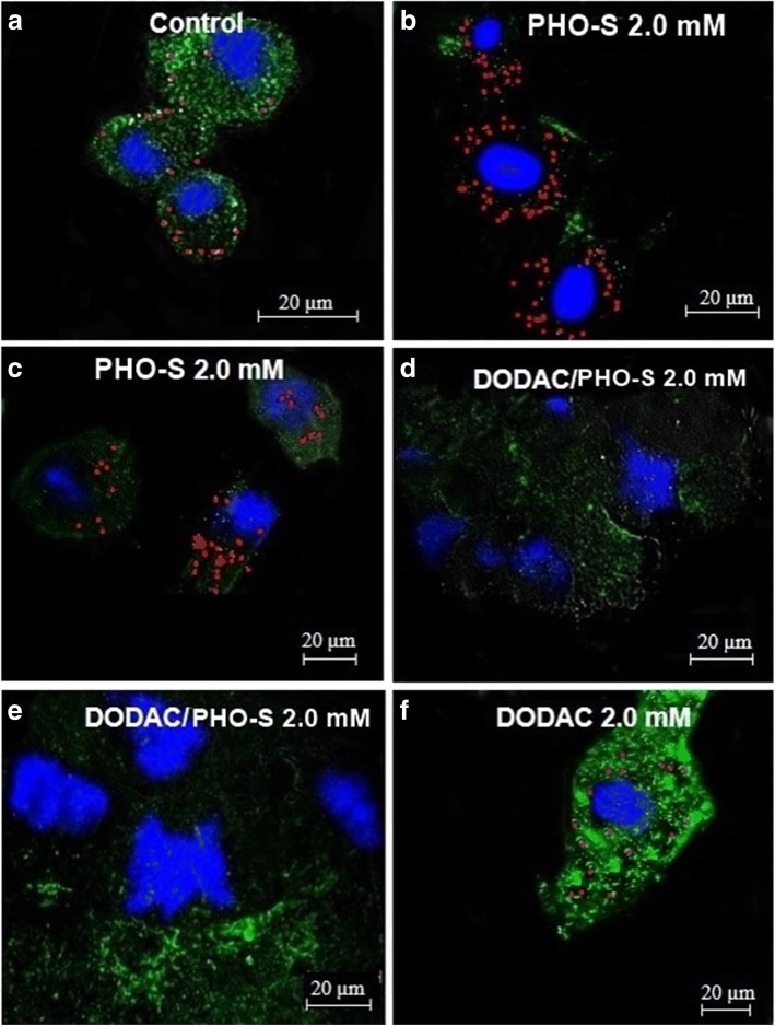
Liposomes (DODAC/PHO-S) were prepared by ultrasonication. The cell cycle phases, protein expression and types of cell's death on Hepa1c1c7 were analyzed by flow cytometry. The internalisation of liposomes, mitochondrial electrical potential and lysosomal stability were also evaluated by confocal laser scanning microscopy.
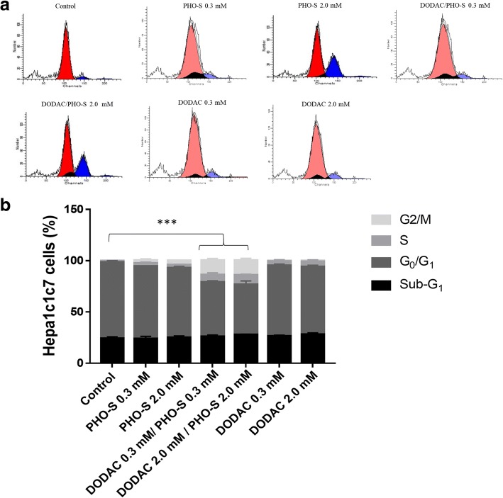
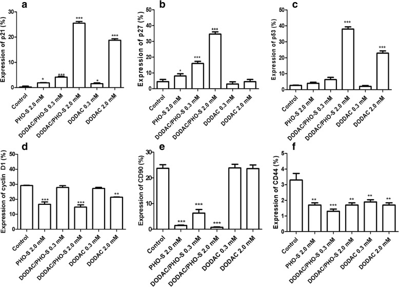
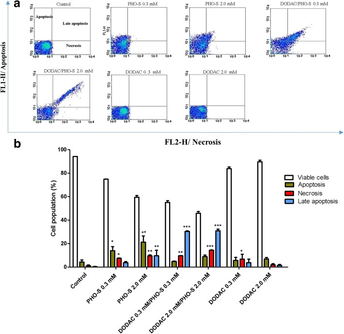
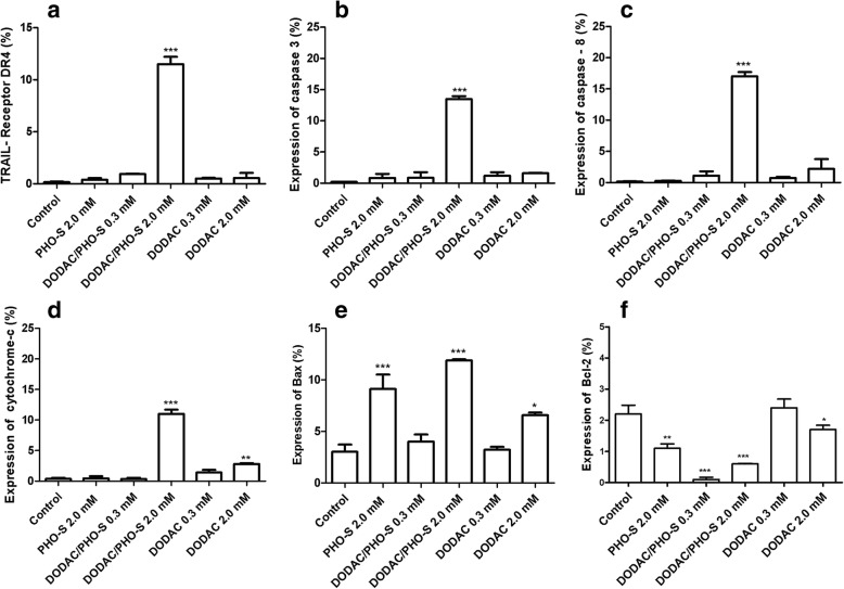
After treatment with liposomes (DODAC/PHO-S), we observed a significant increase in the population of Hepa1c1c7 cells experiencing cell cycle arrest in the S and G/M phases, and this treatment was significantly more effective to promote cell death by apoptosis. There also was a decrease in the mitochondrial electrical potential; changes in the lysosomes; nuclear fragmentation and catastrophic changes in Hepa1c1c7 cells. The liposomes additionally promoted increases in the expression of DR4 receptor, caspases 3 and 8, cytochrome c, p53, p21, p27 and Bax. There was also a decrease in the expression of Bcl-2, cyclin D1, CD90 and CD44 proteins.
The overall results showed that DODAC/PHO-S liposomes were more effective than PHO-S alone, in promoting cytotoxicity Hepa1c1c7 tumor cells, activating the intrinsic and extrinsic pathways of programmed cell death.
目前的研究表明,二辛基二甲基氯化铵/合成磷酸乙醇胺(DODAC/PHO-S)脂质体对Hepa1c1c7和B16F10小鼠肿瘤细胞具有细胞毒性,且比磷酸乙醇胺(PHO-S)的比例更高。因此,我们的目的是评估DODAC/PHO-S脂质体在阐明细胞死亡机制方面的潜力,即与单独的PHO-S相比,该脂质体如何在肝癌Hepa1c1c7细胞中诱导细胞毒性。
通过超声处理制备脂质体(DODAC/PHO-S)。采用流式细胞术分析Hepa1c1c7细胞的细胞周期阶段、蛋白质表达和细胞死亡类型。还通过共聚焦激光扫描显微镜评估脂质体的内化、线粒体膜电位和溶酶体稳定性。
用脂质体(DODAC/PHO-S)处理后,我们观察到处于S期和G/M期细胞周期停滞的Hepa1c1c7细胞群体显著增加,并且这种处理在通过凋亡促进细胞死亡方面显著更有效。线粒体膜电位也降低;溶酶体发生变化;Hepa1c1c7细胞出现核碎裂和灾难性变化。脂质体还促进了死亡受体4(DR4)、半胱天冬酶3和8、细胞色素c、p53、p21、p27和Bax表达的增加。Bcl-2、细胞周期蛋白D1、CD90和CD44蛋白的表达也降低。
总体结果表明,DODAC/PHO-S脂质体在促进Hepa1c1c7肿瘤细胞的细胞毒性、激活程序性细胞死亡的内在和外在途径方面比单独的PHO-S更有效。