• 文献检索
  • 文档翻译
  • 深度研究
  • 学术资讯
  • Suppr Zotero 插件Zotero 插件
  • 邀请有礼
  • 套餐&价格
  • 历史记录
应用&插件
Suppr Zotero 插件Zotero 插件浏览器插件Mac 客户端Windows 客户端微信小程序
定价
高级版会员购买积分包购买API积分包
服务
文献检索文档翻译深度研究API 文档MCP 服务
关于我们
关于 Suppr公司介绍联系我们用户协议隐私条款
关注我们

Suppr 超能文献

核心技术专利:CN118964589B侵权必究
粤ICP备2023148730 号-1Suppr @ 2026

文献检索

告别复杂PubMed语法,用中文像聊天一样搜索,搜遍4000万医学文献。AI智能推荐,让科研检索更轻松。

立即免费搜索

文件翻译

保留排版,准确专业,支持PDF/Word/PPT等文件格式,支持 12+语言互译。

免费翻译文档

深度研究

AI帮你快速写综述,25分钟生成高质量综述,智能提取关键信息,辅助科研写作。

立即免费体验

应用定量形态学和功能学特征评估乳腺动态对比增强磁共振成像中的腋窝淋巴结。

Use of Quantitative Morphological and Functional Features for Assessment of Axillary Lymph Node in Breast Dynamic Contrast-Enhanced Magnetic Resonance Imaging.

机构信息

Radiology Unit, "Dipartimento di Supporto ai Percorsi Oncologici Area Diagnostica, Istituto Nazionale Tumori-IRCCS-Fondazione G. Pascale", Via Mariano Semmola, Naples, Italy.

Department of Electrical Engineering and Information Technologies, University "Federico II" of Naples, Via Claudio, Naples, Italy.

出版信息

Biomed Res Int. 2018 May 30;2018:2610801. doi: 10.1155/2018/2610801. eCollection 2018.

DOI:10.1155/2018/2610801
PMID:30003092
原文链接:https://pmc.ncbi.nlm.nih.gov/articles/PMC5998166/
Abstract

BACKGROUND

Axillary lymph-node assessment is considered one of the most important prognostic factors concerning breast cancer survival.

OBJECTIVE

We investigated the discriminative power of morphological and functional features in assessing the axillary lymph node.

METHODS

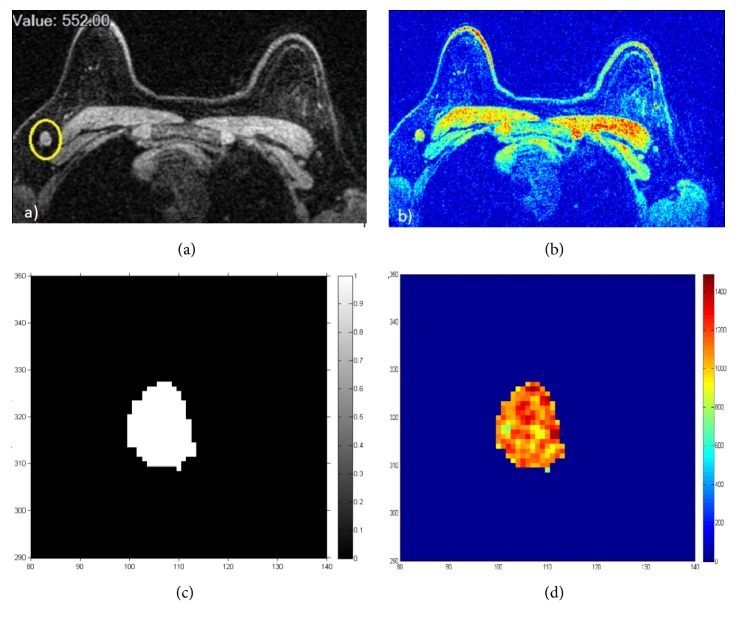
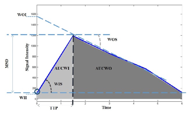
We retrospectively analysed data from 52 consecutive patients who undergone DCE-MRI and were diagnosed with primary breast carcinoma: 94 lymph nodes were identified. Per each lymph node, we extracted morphological features: circularity, compactness, convexity, curvature, elongation, diameter, eccentricity, irregularity, radial length, entropy, rectangularity, roughness, smoothness, sphericity, spiculation, surface, and volume. Moreover, we extracted functional features: time to peak (TTP), maximum signal difference (MSD), wash-in intercept (WII), wash-out intercept (WOI), wash-in slope (WIS), wash-out slope (WOS), area under gadolinium curve (AUGC), area under wash-in (AUWI), and area under wash-out (AUWO). Selection of important features in predicting metastasis has been done by means of receiver operating characteristic (ROC) analysis. Performance of linear discriminant analysis was analysed.

RESULTS

All morphological features but circularity showed a significant difference between median values of metastatic lymph nodes group and nonmetastatic lymph nodes group. All dynamic parameters except for MSD and WOS showed a statistically significant difference between median values of metastatic lymph nodes group and nonmetastatic lymph nodes group. Best results for discrimination of metastatic and nonmetastatic lymph nodes were obtained by AUGC (accuracy 75.8%), WIS (accuracy 71.0%), WOS (accuracy 71.0%), and AUCWO (accuracy 72.6%) for dynamic features and by compactness (accuracy 82.3%), curvature (accuracy 71.0%), radial length (accuracy 71.0%), roughness (accuracy 74.2%), smoothness (accuracy 77.2%), and speculation (accuracy 72.6%) for morphological features. Linear combination of all morphological and/or of all dynamic features did not increase accuracy in metastatic lymph nodes discrimination.

CONCLUSIONS

Compactness as morphological feature and area under time-intensity curve as dynamic feature were the best parameters in identifying metastatic lymph nodes on breast MRI.

摘要

背景

腋窝淋巴结评估被认为是与乳腺癌生存相关的最重要的预后因素之一。

目的

我们研究了形态和功能特征在评估腋窝淋巴结中的鉴别能力。

方法

我们回顾性分析了 52 例连续接受 DCE-MRI 检查并被诊断为原发性乳腺癌的患者的数据:共识别出 94 个淋巴结。对于每个淋巴结,我们提取形态特征:圆形度、紧致度、凸度、曲率、伸长率、直径、偏心率、不规则性、径向长度、熵、矩形度、粗糙度、平滑度、球形度、刺状度、表面和体积。此外,我们还提取了功能特征:达峰时间(TTP)、最大信号差(MSD)、灌洗截距(WII)、洗脱截距(WOI)、灌洗斜率(WIS)、洗脱斜率(WOS)、钆曲线下面积(AUGC)、灌洗下面积(AUWI)和洗脱下面积(AUWO)。通过受试者工作特征(ROC)分析选择预测转移的重要特征。分析了线性判别分析的性能。

结果

除圆形度外,所有形态特征的中位数在转移性淋巴结组和非转移性淋巴结组之间均有显著差异。除 MSD 和 WOS 外,所有动态参数的中位数在转移性淋巴结组和非转移性淋巴结组之间均有统计学差异。用于区分转移性和非转移性淋巴结的最佳结果是通过 AUGC(准确性 75.8%)、WIS(准确性 71.0%)、WOS(准确性 71.0%)和 AUCWO(准确性 72.6%)获得的动态特征,以及通过紧凑度(准确性 82.3%)、曲率(准确性 71.0%)、径向长度(准确性 71.0%)、粗糙度(准确性 74.2%)、平滑度(准确性 77.2%)和推测(准确性 72.6%)获得的形态特征。所有形态和/或所有动态特征的线性组合并不能提高转移性淋巴结鉴别诊断的准确性。

结论

形态学特征中的紧凑度和动力学特征中的时间-强度曲线下面积是在乳腺 MRI 上识别转移性淋巴结的最佳参数。

https://cdn.ncbi.nlm.nih.gov/pmc/blobs/3ddb/5998166/a1ff38b443d7/BMRI2018-2610801.002.jpg
https://cdn.ncbi.nlm.nih.gov/pmc/blobs/3ddb/5998166/3c088c71effa/BMRI2018-2610801.001.jpg
https://cdn.ncbi.nlm.nih.gov/pmc/blobs/3ddb/5998166/a1ff38b443d7/BMRI2018-2610801.002.jpg
https://cdn.ncbi.nlm.nih.gov/pmc/blobs/3ddb/5998166/3c088c71effa/BMRI2018-2610801.001.jpg
https://cdn.ncbi.nlm.nih.gov/pmc/blobs/3ddb/5998166/a1ff38b443d7/BMRI2018-2610801.002.jpg

相似文献

1
Use of Quantitative Morphological and Functional Features for Assessment of Axillary Lymph Node in Breast Dynamic Contrast-Enhanced Magnetic Resonance Imaging.应用定量形态学和功能学特征评估乳腺动态对比增强磁共振成像中的腋窝淋巴结。
Biomed Res Int. 2018 May 30;2018:2610801. doi: 10.1155/2018/2610801. eCollection 2018.
2
In vitro diagnosis of axillary lymph node metastases in breast cancer by spectrum analysis of radio frequency echo signals.通过射频回波信号频谱分析对乳腺癌腋窝淋巴结转移进行体外诊断。
Ultrasound Med Biol. 1998 Oct;24(8):1151-9. doi: 10.1016/s0301-5629(98)00100-8.
3
Role of diffusion-weighted MRI: predicting axillary lymph node metastases in breast cancer.扩散加权磁共振成像的作用:预测乳腺癌腋窝淋巴结转移
Acta Radiol. 2014 Oct;55(8):909-16. doi: 10.1177/0284185113509094. Epub 2013 Nov 14.
4
Assessment of axillary lymph nodes in patients with breast cancer with diffusion-weighted MR imaging in combination with routine and dynamic contrast MR imaging.采用扩散加权磁共振成像联合常规及动态对比增强磁共振成像评估乳腺癌患者腋窝淋巴结。
Breast Cancer. 2016 May;23(3):525-32. doi: 10.1007/s12282-015-0598-7. Epub 2015 Mar 13.
5
Adding contrast-enhanced ultrasound markers to conventional axillary ultrasound improves specificity for predicting axillary lymph node metastasis in patients with breast cancer.在常规腋窝超声中添加增强超声标志物可提高预测乳腺癌患者腋窝淋巴结转移的特异性。
Br J Radiol. 2021 Feb 1;94(1118):20200874. doi: 10.1259/bjr.20200874. Epub 2020 Dec 22.
6
Differentiating axillary lymph node metastasis in invasive breast cancer patients: A comparison of radiomic signatures from multiparametric breast MR sequences.鉴别浸润性乳腺癌患者腋窝淋巴结转移:多参数乳腺磁共振序列的放射组学特征比较。
J Magn Reson Imaging. 2019 Oct;50(4):1125-1132. doi: 10.1002/jmri.26701. Epub 2019 Mar 7.
7
Dual-modal computer-assisted evaluation of axillary lymph node metastasis in breast cancer patients on both real-time elastography and B-mode ultrasound.实时超声弹性成像与 B 型超声双模计算机辅助评估乳腺癌患者腋窝淋巴结转移
Eur J Radiol. 2017 Oct;95:66-74. doi: 10.1016/j.ejrad.2017.07.027. Epub 2017 Aug 1.
8
Preoperative MRI improves prediction of extensive occult axillary lymph node metastases in breast cancer patients with a positive sentinel lymph node biopsy.术前 MRI 可提高前哨淋巴结活检阳性乳腺癌患者隐匿性腋窝广泛淋巴结转移的预测能力。
Acad Radiol. 2014 Jan;21(1):92-8. doi: 10.1016/j.acra.2013.10.001.
9
Radiomic features of axillary lymph nodes based on pharmacokinetic modeling DCE-MRI allow preoperative diagnosis of their metastatic status in breast cancer.基于药代动力学模型 DCE-MRI 的腋窝淋巴结放射组学特征可实现乳腺癌腋窝淋巴结转移状态的术前诊断。
PLoS One. 2021 Mar 1;16(3):e0247074. doi: 10.1371/journal.pone.0247074. eCollection 2021.
10
Correlation between the contrast-enhanced ultrasound image features and axillary lymph node metastasis of primary breast cancer and its diagnostic value.原发性乳腺癌超声造影图像特征与腋窝淋巴结转移的相关性及其诊断价值。
Clin Transl Oncol. 2021 Jan;23(1):155-163. doi: 10.1007/s12094-020-02407-6. Epub 2020 Jun 1.

引用本文的文献

1
Integrating Radiogenomics and Machine Learning in Musculoskeletal Oncology Care.将放射基因组学与机器学习整合于肌肉骨骼肿瘤护理中。
Diagnostics (Basel). 2025 May 29;15(11):1377. doi: 10.3390/diagnostics15111377.
2
Application of CT and MRI images based on an artificial intelligence algorithm for predicting lymph node metastasis in breast cancer patients: a meta-analysis.基于人工智能算法的 CT 和 MRI 图像在预测乳腺癌患者淋巴结转移中的应用:一项荟萃分析。
BMC Cancer. 2023 Nov 22;23(1):1134. doi: 10.1186/s12885-023-11638-z.
3
Recent Advances in Ultrasound Breast Imaging: From Industry to Clinical Practice.

本文引用的文献

1
Pattern Recognition Approaches for Breast Cancer DCE-MRI Classification: A Systematic Review.乳腺癌动态对比增强磁共振成像分类的模式识别方法:一项系统综述。
J Med Biol Eng. 2016;36(4):449-459. doi: 10.1007/s40846-016-0163-7. Epub 2016 Aug 31.
2
Potential Role of PET/MRI for Imaging Metastatic Lymph Nodes in Head and Neck Cancer.PET/MRI在头颈部癌转移淋巴结成像中的潜在作用
AJR Am J Roentgenol. 2016 Aug;207(2):248-56. doi: 10.2214/AJR.16.16265. Epub 2016 May 10.
3
Integration of DCE-MRI and DW-MRI Quantitative Parameters for Breast Lesion Classification.
超声乳腺成像的最新进展:从工业到临床实践
Diagnostics (Basel). 2023 Mar 4;13(5):980. doi: 10.3390/diagnostics13050980.
4
Clinical prediction model based on 18F-FDG PET/CT plus contrast-enhanced MRI for axillary lymph node macrometastasis.基于18F-FDG PET/CT联合对比增强MRI的腋窝淋巴结大转移临床预测模型
Front Oncol. 2022 Sep 13;12:989650. doi: 10.3389/fonc.2022.989650. eCollection 2022.
5
A Novel Combined Nomogram Model for Predicting the Pathological Complete Response to Neoadjuvant Chemotherapy in Invasive Breast Carcinoma of No Specific Type: Real-World Study.一种预测非特殊类型浸润性乳腺癌新辅助化疗病理完全缓解的新型联合列线图模型:真实世界研究
Front Oncol. 2022 Jun 6;12:916526. doi: 10.3389/fonc.2022.916526. eCollection 2022.
6
Not only lymphadenopathy: case of chest lymphangitis assessed with MRI after COVID 19 vaccine.不仅仅是淋巴结病:19 岁后接种新冠疫苗后经 MRI 评估的胸部淋巴管炎病例 。 你提供的原文中“COVID 19 vaccine”表述有误,应该是“COVID-19 vaccine”,正确译文如下: 不仅仅是淋巴结病:接种新冠疫苗后经 MRI 评估的胸部淋巴管炎病例 。
Infect Agent Cancer. 2022 Mar 17;17(1):8. doi: 10.1186/s13027-022-00419-1.
7
A meta-analysis of the diagnostic performance of machine learning-based MRI in the prediction of axillary lymph node metastasis in breast cancer patients.基于机器学习的MRI对乳腺癌患者腋窝淋巴结转移预测诊断性能的荟萃分析。
Insights Imaging. 2021 Nov 3;12(1):156. doi: 10.1186/s13244-021-01034-1.
8
A prospective study of dual-energy computed tomography for differentiating metastatic and non-metastatic lymph nodes of colorectal cancer.双能计算机断层扫描鉴别结直肠癌转移与非转移淋巴结的前瞻性研究。
Quant Imaging Med Surg. 2021 Aug;11(8):3448-3459. doi: 10.21037/qims-20-3.
9
Non-invasive prediction of lymph node status for patients with early-stage invasive breast cancer based on a morphological feature from ultrasound images.基于超声图像形态学特征对早期浸润性乳腺癌患者淋巴结状态的无创预测
Quant Imaging Med Surg. 2021 Aug;11(8):3399-3407. doi: 10.21037/qims-20-1201.
10
Radiomic features of axillary lymph nodes based on pharmacokinetic modeling DCE-MRI allow preoperative diagnosis of their metastatic status in breast cancer.基于药代动力学模型 DCE-MRI 的腋窝淋巴结放射组学特征可实现乳腺癌腋窝淋巴结转移状态的术前诊断。
PLoS One. 2021 Mar 1;16(3):e0247074. doi: 10.1371/journal.pone.0247074. eCollection 2021.
用于乳腺病变分类的动态对比增强磁共振成像(DCE-MRI)和扩散加权磁共振成像(DW-MRI)定量参数整合
Biomed Res Int. 2015;2015:237863. doi: 10.1155/2015/237863. Epub 2015 Aug 3.
4
Using quantitative image analysis to classify axillary lymph nodes on breast MRI: a new application for the Z 0011 Era.利用定量图像分析在乳腺磁共振成像上对腋窝淋巴结进行分类:Z 0011时代的一项新应用。
Eur J Radiol. 2015 Mar;84(3):392-397. doi: 10.1016/j.ejrad.2014.12.003. Epub 2014 Dec 15.
5
The Comparative Study of Ultrasonography, Contrast-Enhanced MRI, and (18)F-FDG PET/CT for Detecting Axillary Lymph Node Metastasis in T1 Breast Cancer.超声、增强 MRI 和 (18)F-FDG PET/CT 对 T1 期乳腺癌腋窝淋巴结转移的对比研究。
J Breast Cancer. 2013 Sep;16(3):315-21. doi: 10.4048/jbc.2013.16.3.315. Epub 2013 Sep 30.
6
A new, preoperative, MRI-based scoring system for diagnosing malignant axillary lymph nodes in women evaluated for breast cancer.一种新的、术前基于 MRI 的评分系统,用于诊断女性乳腺癌评估中的恶性腋窝淋巴结。
Eur J Radiol. 2012 Oct;81(10):2602-12. doi: 10.1016/j.ejrad.2012.03.019. Epub 2012 Apr 21.
7
Accuracy of predicting axillary lymph node positivity by physical examination, mammography, ultrasonography, and magnetic resonance imaging.体格检查、乳房 X 线摄影术、超声检查和磁共振成像预测腋窝淋巴结阳性的准确性。
Ann Surg Oncol. 2012 Jun;19(6):1825-30. doi: 10.1245/s10434-011-2200-7. Epub 2012 Jan 7.
8
Application of MR mammography beyond local staging: is there a potential to accurately assess axillary lymph nodes? evaluation of an extended protocol in an initial prospective study.MR 乳腺摄影在局部分期之外的应用:是否有可能准确评估腋窝淋巴结?一项扩展方案在初步前瞻性研究中的评估。
AJR Am J Roentgenol. 2011 May;196(5):W641-7. doi: 10.2214/AJR.10.4889.
9
Positron emission tomography (PET) and magnetic resonance imaging (MRI) for the assessment of axillary lymph node metastases in early breast cancer: systematic review and economic evaluation.正电子发射断层扫描(PET)和磁共振成像(MRI)在早期乳腺癌腋窝淋巴结转移评估中的应用:系统评价和经济评估。
Health Technol Assess. 2011 Jan;15(4):iii-iv, 1-134. doi: 10.3310/hta15040.
10
An expectation-maximisation approach for simultaneous pixel classification and tracer kinetic modelling in dynamic contrast enhanced-magnetic resonance imaging.一种用于动态对比增强磁共振成像中同时进行像素分类和示踪剂动力学建模的期望最大化方法。
Med Biol Eng Comput. 2011 Apr;49(4):485-95. doi: 10.1007/s11517-010-0695-x. Epub 2010 Nov 3.