Department of Radiology, First Hospital of China Medical University, Shenyang, Liaoning Province, China.
Sino-Dutch Biomedical and Infornation Engineering School, Northeastern University, Shenyang, Liaoning Province, China.
J Magn Reson Imaging. 2019 Oct;50(4):1125-1132. doi: 10.1002/jmri.26701. Epub 2019 Mar 7.
The axillary lymph node status is critical for breast cancer staging and individualized treatment planning.
To assess the effect of determining axillary lymph node (ALN) metastasis by breast MRI-derived radiomic signatures, and compare the discriminating abilities of different MR sequences.
Retrospective.
In all, 120 breast cancer patients, 59 with ALN metastasis and 61 without metastasis, all confirmed by pathology.
FIELD STRENGTH/SEQUENCE: 3 .0T scanner with T -weighted imaging, T -weighted imaging, diffusion-weighted imaging, and dynamic contrast-enhanced (DCE) sequences.
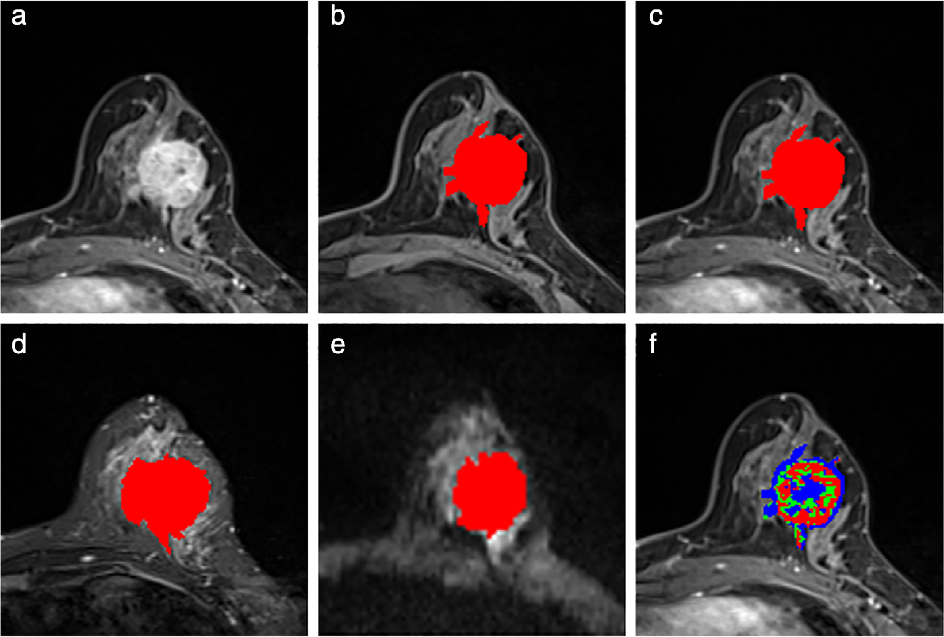
Typical morphological and texture features of the segmented tumor were extracted from four sequences, ie, T WI, T WI, DWI, and the second postcontrast phase (CE2) of the dynamic contrast-enhanced sequences. Additional contrast enhancement kinetic features were extracted from all DCE sequences (one pre- and seven postcontrast phases). Linear discriminant analysis classifiers were built and compared when using features from an individual sequence or the combination of the sequences in differentiating the ALN metastasis status.
Mann-Whitney U-test, Fisher's exact test, least absolute shrinkage selection operator (LASSO) regression, and receiver operating characteristic analysis were performed.
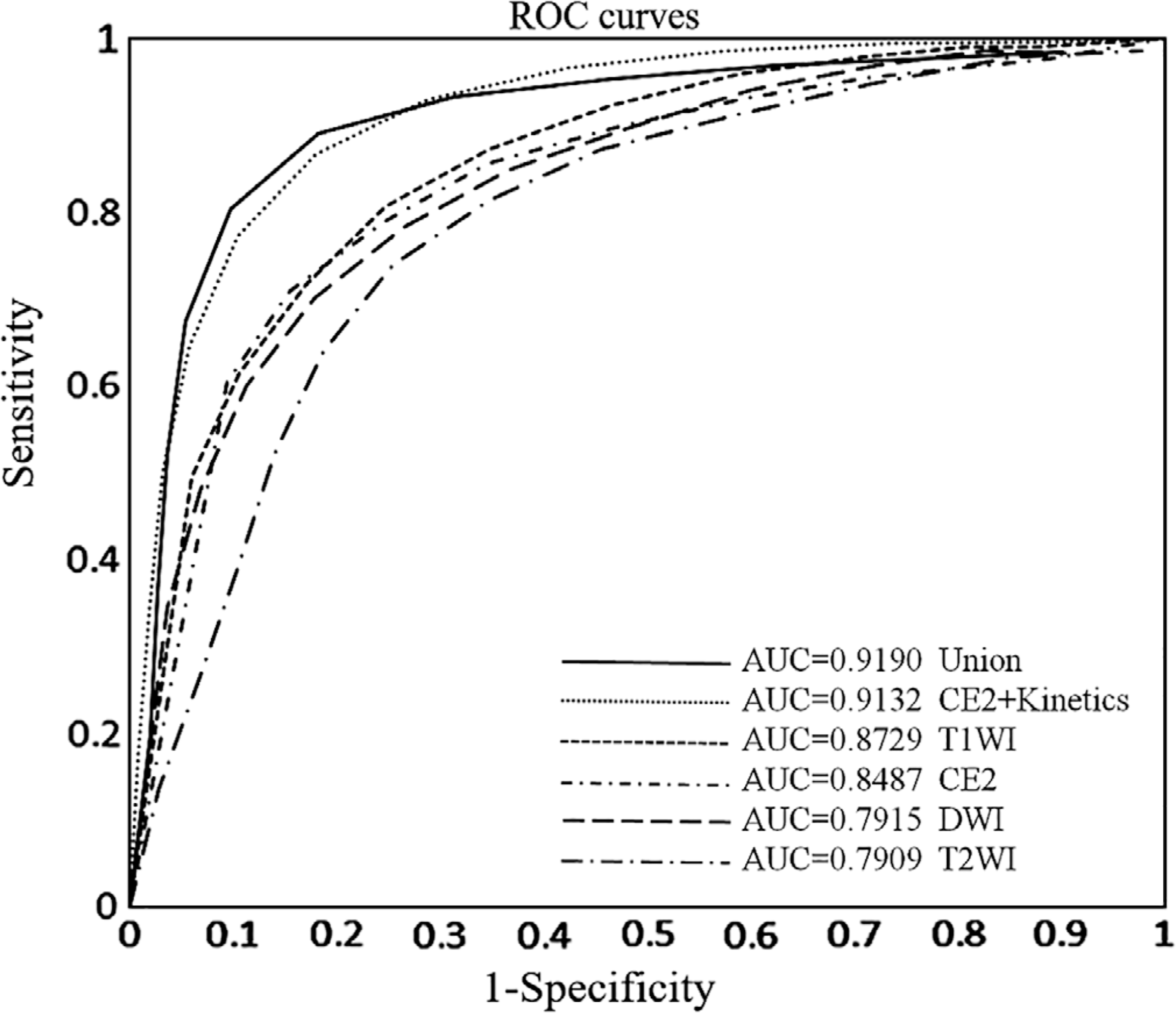
The accuracy/AUC of the four sequences was 79%/0.87, 77%/0.85, 74%/0.79, and 79%/0.85 for the T WI, CE2, T WI, and DWI, respectively. When CE2 was augmented by adding kinetic features, the model achieved the highest performance (accuracy = 0.86 and AUC = 0.91). When all features from the four sequences and the kinetics were combined, it did not lead to a further increase in the performance (P = 0.48).
Breast tumor's radiomic signatures from preoperative breast MRI sequences are associated with the ALN metastasis status, where CE2 phase and the contrast enhancement kinetic features lead to the highest classification effect. Level of Evidence 3 Technical Efficacy Stage 2 J. Magn. Reson. Imaging 2019;50:1125-1132.
腋窝淋巴结状态对乳腺癌分期和个体化治疗计划至关重要。
评估通过乳腺 MRI 衍生的放射组学特征确定腋窝淋巴结 (ALN) 转移的效果,并比较不同 MR 序列的区分能力。
回顾性。
共 120 例乳腺癌患者,59 例腋窝淋巴结转移,61 例无转移,均经病理证实。
磁场强度/序列:3.0T 扫描仪,T1 加权成像、T2 加权成像、扩散加权成像和动态对比增强(DCE)序列。
从四个序列(T1WI、T2WI、DWI 和 DCE 序列的第二期对比增强)提取分割肿瘤的典型形态和纹理特征。从所有 DCE 序列(一期和七期对比后)提取额外的对比增强动力学特征。当使用单个序列或序列组合的特征来区分 ALN 转移状态时,构建并比较线性判别分析分类器。
采用 Mann-Whitney U 检验、Fisher 精确检验、最小绝对收缩和选择算子(LASSO)回归和受试者工作特征分析。
T1WI、CE2、T2WI 和 DWI 的准确率/ AUC 分别为 79%/0.87、77%/0.85、74%/0.79 和 79%/0.85。当在 CE2 期添加动力学特征时,模型的性能最高(准确率=0.86,AUC=0.91)。当将四个序列和动力学的所有特征结合起来时,性能并没有进一步提高(P=0.48)。
术前乳腺 MRI 序列的乳腺肿瘤放射组学特征与 ALN 转移状态相关,CE2 期和对比增强动力学特征可产生最高的分类效果。
证据水平 3 技术功效 2 级
磁共振成像杂志 2019;50:1125-1132