Skowerski M, Wozniak-Skowerska I, Hoffmann A, Nowak S, Skowerski T, Sosnowski M, Wnuk-Wojnar A M, Mizia-Stec K
Department of Cardiology, School of Health Sciences, Medical University of Silesia, Katowice, Poland.
First Department of Cardiology, School of Medicine in Katowice, Medical University of Silesia, Katowice, Poland.
BMC Cardiovasc Disord. 2018 Jul 13;18(1):146. doi: 10.1186/s12872-018-0884-3.
It has been suggested that changes in pulmonary veins (PV) and left atrium (LA) anatomy may have an influence on initiating atrial fibrillation (AF) and the effectiveness of pulmonary vein isolation (PVI) in patients (pts) with atrial fibrillation. The aim of the study was to assess anatomy abnormalities of the PV and LA in the patients with the history of AF and compare it with the control group(CG).
The multi-slice tomography (MSCT) scans were performed in 224 AF pts. before PVI (129 males, mean age 59 ± 9 yrs). The CG consisted of 40 pts. without AF (26 males, age 45 ± 9 yrs). LA and PV anatomy were evaluated. Diameters of PV ostia were measured in two directions: anterior-posterior (AP) and superior-inferior (SI) automatically using Vitrea 4.0.
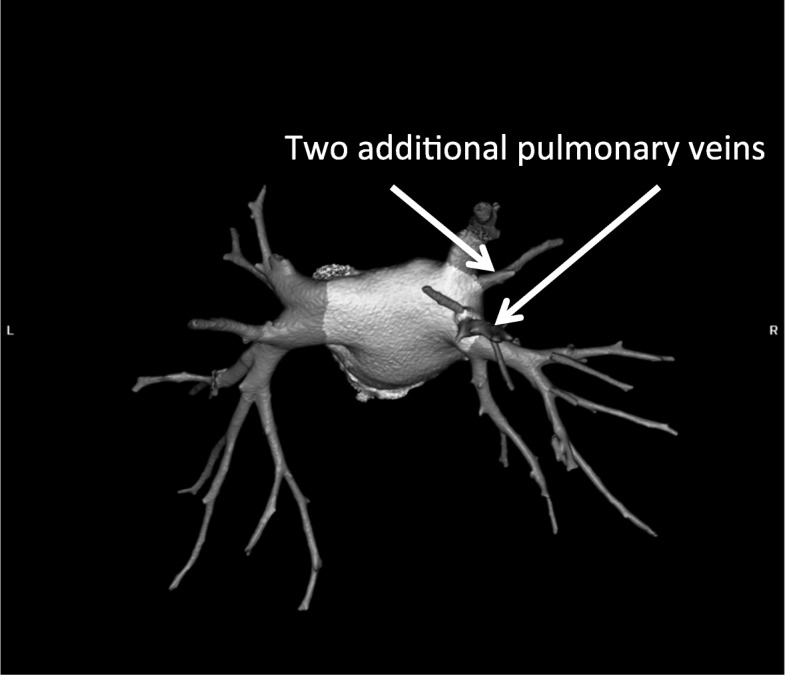
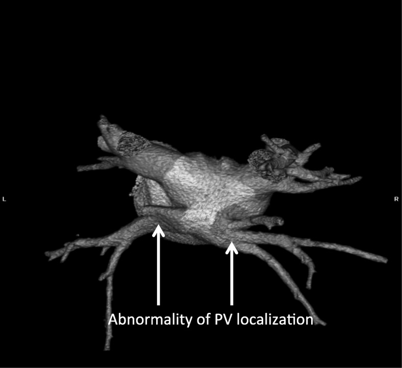
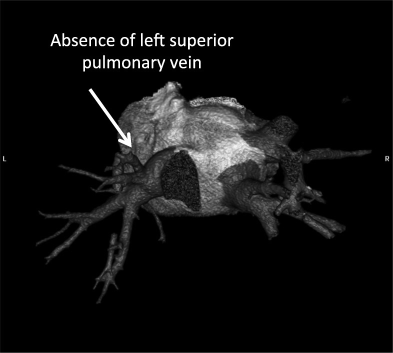
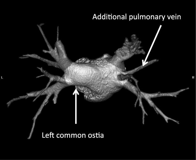
Pulmonary veins anatomy variants were observed more frequently in the atrial fibrillation group - 83 pts. (37%) vs 6 pts. (15%) in CG; 9% (21 pts) left common ostia (CO), 2% (5 pts) right CO, 19% (42 pts) additional right PV (APV), (1.8%) 4 pts. APV left, 8% right early branching (EB) and 3.5% left EB. The LA diameter differed significantly in AF vs CG group (41.2 ± 6 mm vs 35 ± 4.2 mm, p < 0.0001) respectively.
The anomalies of pulmonary vein anatomy occurred more often in pts. with AF. They can be defined as an image biomarkers of atrial fibrillation. Right additional (middle) pulmonary vein was the most important anomaly detected in AF patients as well as enlargered diameters of the LA and PV ostia.
有人提出,肺静脉(PV)和左心房(LA)解剖结构的改变可能会影响房颤(AF)的发生以及房颤患者肺静脉隔离(PVI)的疗效。本研究的目的是评估有房颤病史患者的PV和LA解剖异常情况,并与对照组(CG)进行比较。
对224例房颤患者在进行PVI之前进行多层螺旋CT(MSCT)扫描(129例男性,平均年龄59±9岁)。对照组由40例无房颤患者组成(26例男性,年龄45±9岁)。评估LA和PV的解剖结构。使用Vitrea 4.0自动在前后(AP)和上下(SI)两个方向测量PV开口直径。
房颤组肺静脉解剖变异的观察频率更高——83例(37%),而对照组为6例(15%);9%(21例)为左共同开口(CO),2%(5例)为右CO,19%(42例)为额外右肺静脉(APV),1.8%(4例)为左APV,8%为右早期分支(EB),3.5%为左EB。房颤组与对照组的LA直径有显著差异(分别为41.2±6mm和35±4.2mm,p<0.0001)。
肺静脉解剖异常在房颤患者中更常见。它们可被定义为房颤的影像生物标志物。右额外(中间)肺静脉是在房颤患者中检测到的最重要异常,以及LA和PV开口直径增大。