Developmental and Regenerative Neurobiology, Department of Experimental Medical Science, Lund University, Lund, Sweden.
Lund Stem Cell Center, Lund University, Lund, Sweden.
J Comp Neurol. 2018 Sep 1;526(13):2133-2146. doi: 10.1002/cne.24500. Epub 2018 Jul 31.
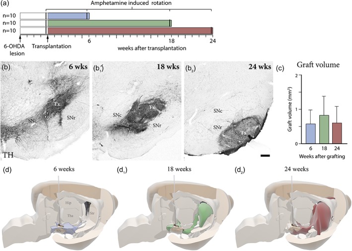
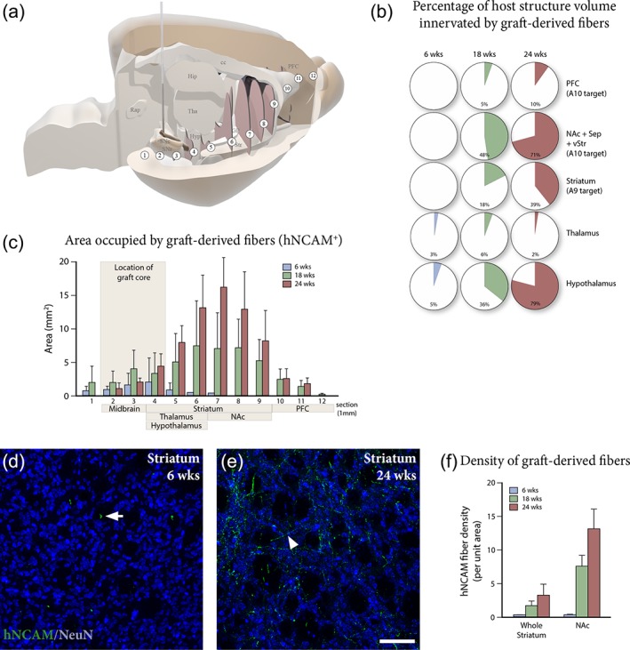
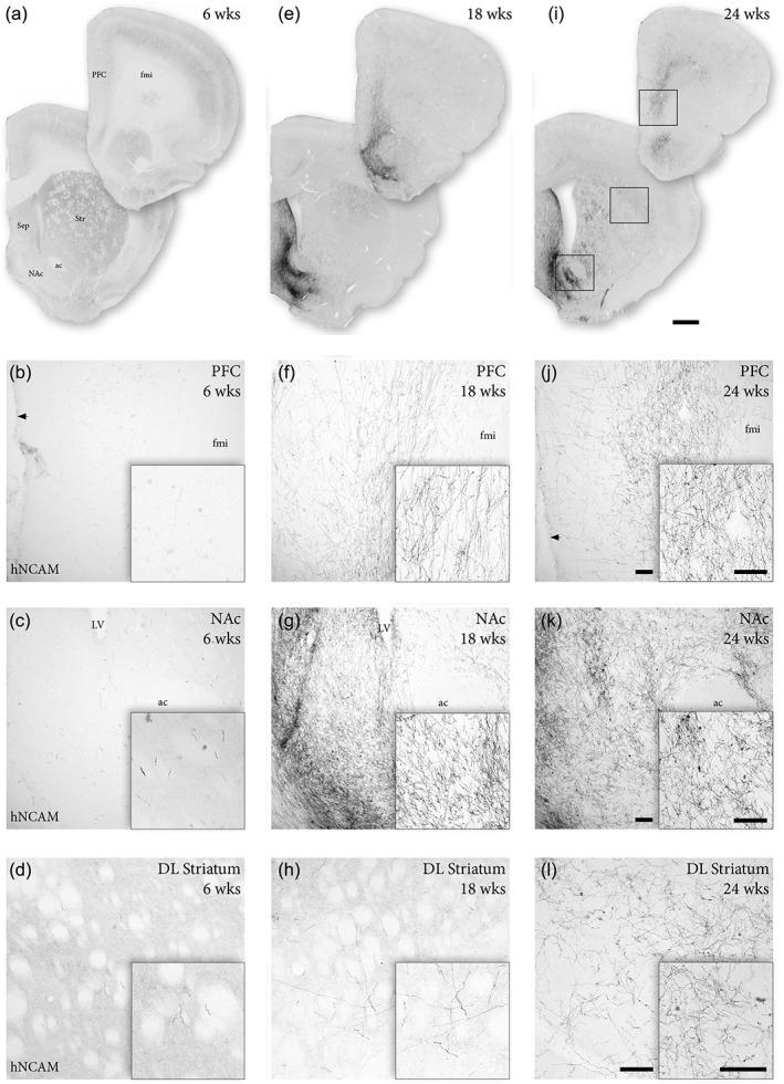
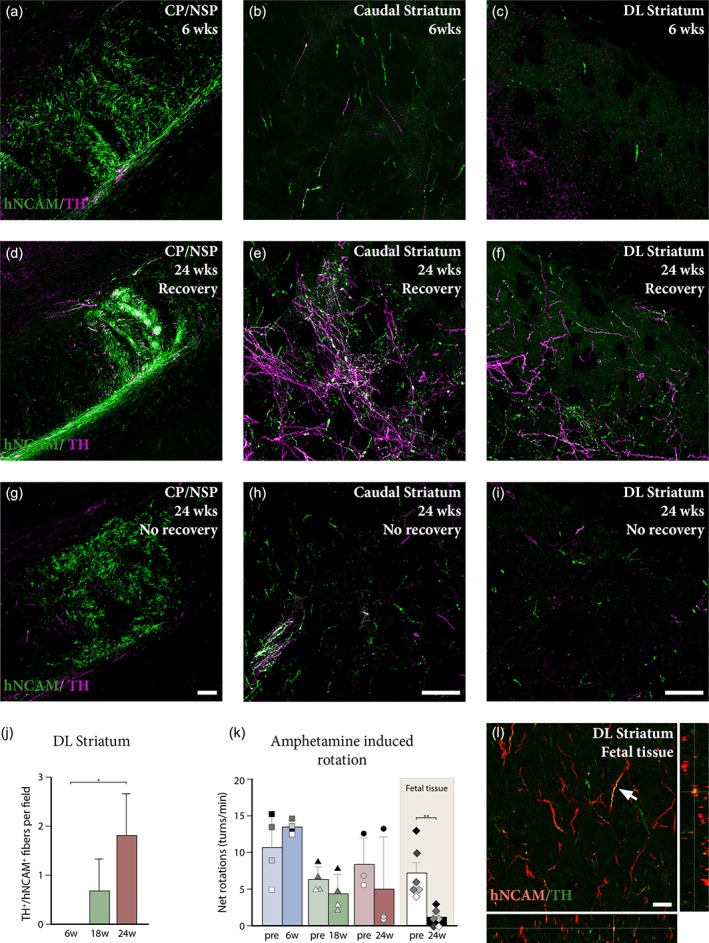
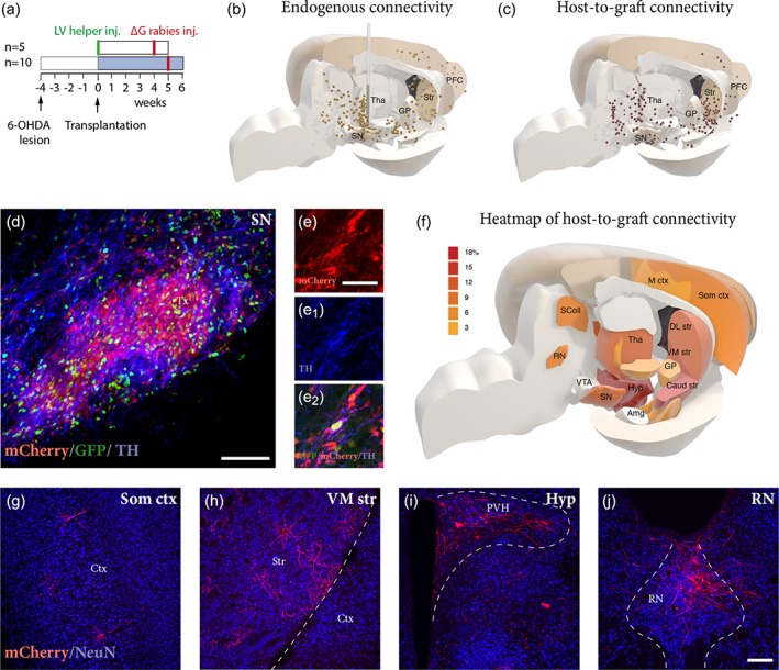
Dopamine (DA) neurons derived from human embryonic stem cells (hESCs) are a promising unlimited source of cells for cell replacement therapy in Parkinson's disease (PD). A number of studies have demonstrated functionality of DA neurons originating from hESCs when grafted to the striatum of rodent and non-human primate models of PD. However, several questions remain in regard to their axonal outgrowth potential and capacity to integrate into host circuitry. Here, ventral midbrain (VM) patterned hESC-derived progenitors were grafted into the midbrain of 6-hydroxydopamine-lesioned rats, and analyzed at 6, 18, and 24 weeks for a time-course evaluation of specificity and extent of graft-derived fiber outgrowth as well as potential for functional recovery. To investigate synaptic integration of the transplanted cells, we used rabies-based monosynaptic tracing to reveal the origin and extent of host presynaptic inputs to grafts at 6 weeks. The results reveal the capacity of grafted neurons to extend axonal projections toward appropriate forebrain target structures progressively over 24 weeks. The timing and extent of graft-derived dopaminergic fibers innervating the dorsolateral striatum matched reduction in amphetamine-induced rotational asymmetry in the animals where recovery could be observed. Monosynaptic tracing demonstrated that grafted cells integrate with host circuitry 6 weeks after transplantation, in a manner that is comparable with endogenous midbrain connectivity. Thus, we demonstrate that VM patterned hESC-derived progenitors grafted to midbrain have the capacity to extensively innervate appropriate forebrain targets, integrate into the host circuitry and that functional recovery can be achieved when grafting fetal or hESC-derived DA neurons to the midbrain.
源自人胚胎干细胞(hESC)的多巴胺(DA)神经元是帕金森病(PD)细胞替代治疗中具有广阔应用前景的细胞来源。大量研究已经证明了源自 hESC 的 DA 神经元在移植到 PD 啮齿动物和非人类灵长类动物模型的纹状体后具有功能。然而,在其轴突生长潜力和与宿主回路整合的能力方面,仍存在一些问题。在这里,腹侧中脑(VM)模式化的 hESC 衍生祖细胞被移植到 6-羟多巴胺损伤大鼠的中脑,在 6、18 和 24 周进行分析,以对移植衍生纤维生长的特异性和程度以及功能恢复的潜力进行时间过程评估。为了研究移植细胞的突触整合,我们使用基于狂犬病毒的单突触示踪来揭示宿主对移植物的突触前输入的起源和程度,这是在 6 周时进行的。结果显示,移植神经元有能力在 24 周内逐渐向适当的前脑靶结构延伸轴突投射。移植多巴胺能纤维在背外侧纹状体的支配程度与动物中可观察到的安非他命诱导的旋转不对称性减少相匹配,而这种减少是可以恢复的。单突触示踪表明,移植后 6 周,移植细胞与宿主回路整合,这种方式与内源性中脑连接方式相当。因此,我们证明了移植到中脑的 VM 模式化的 hESC 衍生祖细胞具有广泛支配适当前脑靶标的能力,与宿主回路整合,并在将胎儿或 hESC 衍生的 DA 神经元移植到中脑时可以实现功能恢复。